P10) Palpable abdominal mass and unexplained weight loss
Review the Learning Outcomes, Hx, PE and Labs, and begin the module with your Provisional Diagnosis. Keep hitting "Next" to move through the module.
Learning Outcomes
- Articulate your relationship with the consulting diagnostic radiologists in the evaluation of a pediatric patient with a palpable abdominal mass.
- Review the DDx considerations in a pediatric patient with a palpable abdominal mass.
- Identify the spectrum of imaging findings in appropriate modalities for evaluating a pediatric patient with a palpable abdominal mass.
History
Physical Exam
Labs
Provisional Diagnosis
Potential Acuity
What is your assessment of the likely acuity for this patient?
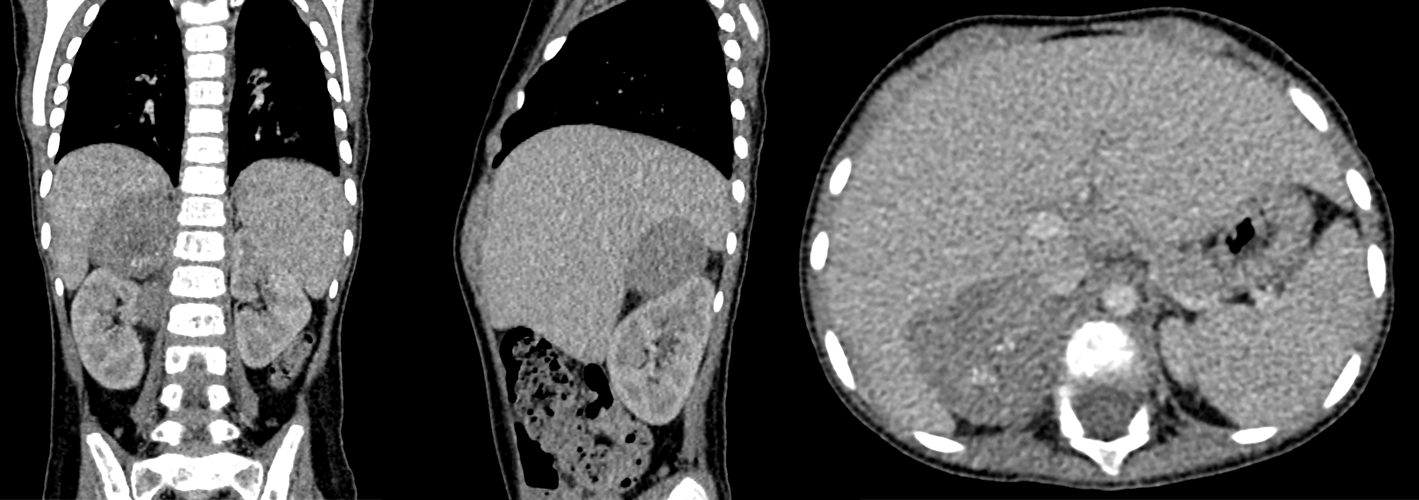
First Imaging Study
What is the first imaging study you will order?
Pertinent Imaging Observations
Click on the links below to view images from the study, and assess these key findings as best you can.

Watch our video
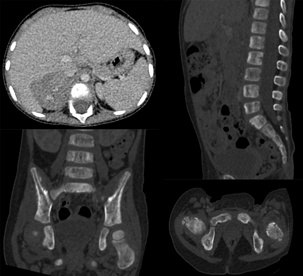
Second Imaging Study
What is the next imaging study you will order?
Pertinent Imaging Observations
Click on the links below to view images from the study, and assess these key findings as best you can.
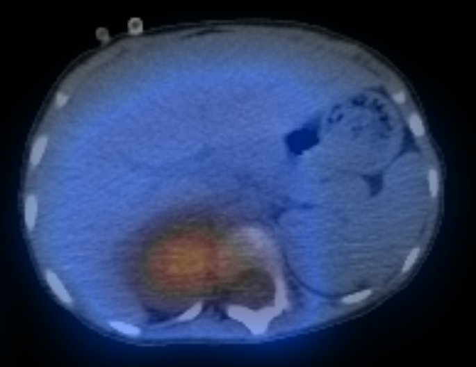
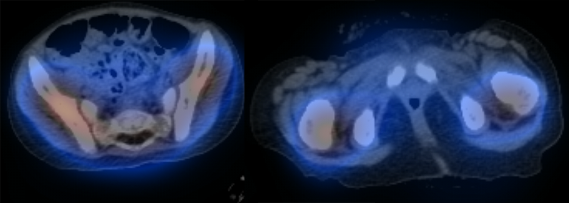
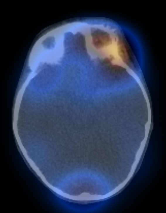
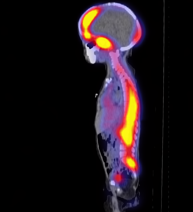
MIBG scan
Increased radiotracer uptake at the primary tumor site is noted in:
Metastasis to the hips and pelvis is:
Metastasis into this area leads to which physical exam finding?
Is there metastasis on the spine?
Watch our video
Third Imaging Study
What is the next imaging study you will order?
What is your Diagnosis now that you have seen the imaging results?
Current Acuity
Initially, you selected and we suggested acuity.
Has your concern for this patient changed?
Assessment and Plan
Please provide your assessment and plan for this patient
Lessons Learned:
- Neuroblastomas are neoplasms originating from neural crest cells. Commonly diagnosed around 1-2 years of age, they are a prevalent childhood cancer and can manifest anywhere along the sympathetic chain.
- Elevated urinary catecholamines, specifically Vanillylmandelic Acid (VMA) and Homovanillic Acid (HVA), are frequently associated with neuroblastoma due to the tumor's increased catecholamine metabolism.
- The clinical presentation of neuroblastoma includes constipation, an abdominal mass palpable across the midline, and opsoclonus-myoclonus ataxia syndrome, colloquially known as "dancing hands and feet".
- Diagnostic modalities, like CT and MIBG imaging, can identify tumor localization, and reveal potential metastatic spread to bones and other organs.
- Histological examination typically reveals small, round blue cells with hyperchromatic nuclei and characteristic Homer Wright rosettes.
- Urinalysis often presents elevated metabolites of catecholamines, specifically homovanillic acid (HVA) and vanillylmandelic acid (VMA).
- Treatment strategies are determined by the staging criteria and the presence of MYCN gene amplification, a marker associated with an increased risk of metastasis.
That's the end of the module! Once you've reviewed the video(s), you can click here for another case challenge.
