N14) Headache, weakness, and unintentional weight loss
Review the Learning Outcomes, Hx, PE and Labs, and begin the module with your Provisional Diagnosis. Keep hitting "Next" to move through the module.
Learning Outcomes
- Articulate your relationship with the consulting diagnostic radiologists in the evaluation of a patient with headache and vomiting.
- Review the DDx considerations in a patient with headache and vomiting.
- Identify the spectrum of imaging findings in appropriate modalities for evaluating patients with headache and vomiting.
History
Physical Exam
Labs
Provisional Diagnosis
Potential Acuity
What is your assessment of the likely acuity for this patient?
First Imaging Study
What is the first imaging study you will order?
Pertinent Imaging Observations
Click on the links below to view images from the study, and assess these key findings as best you can.
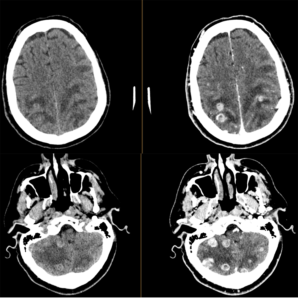
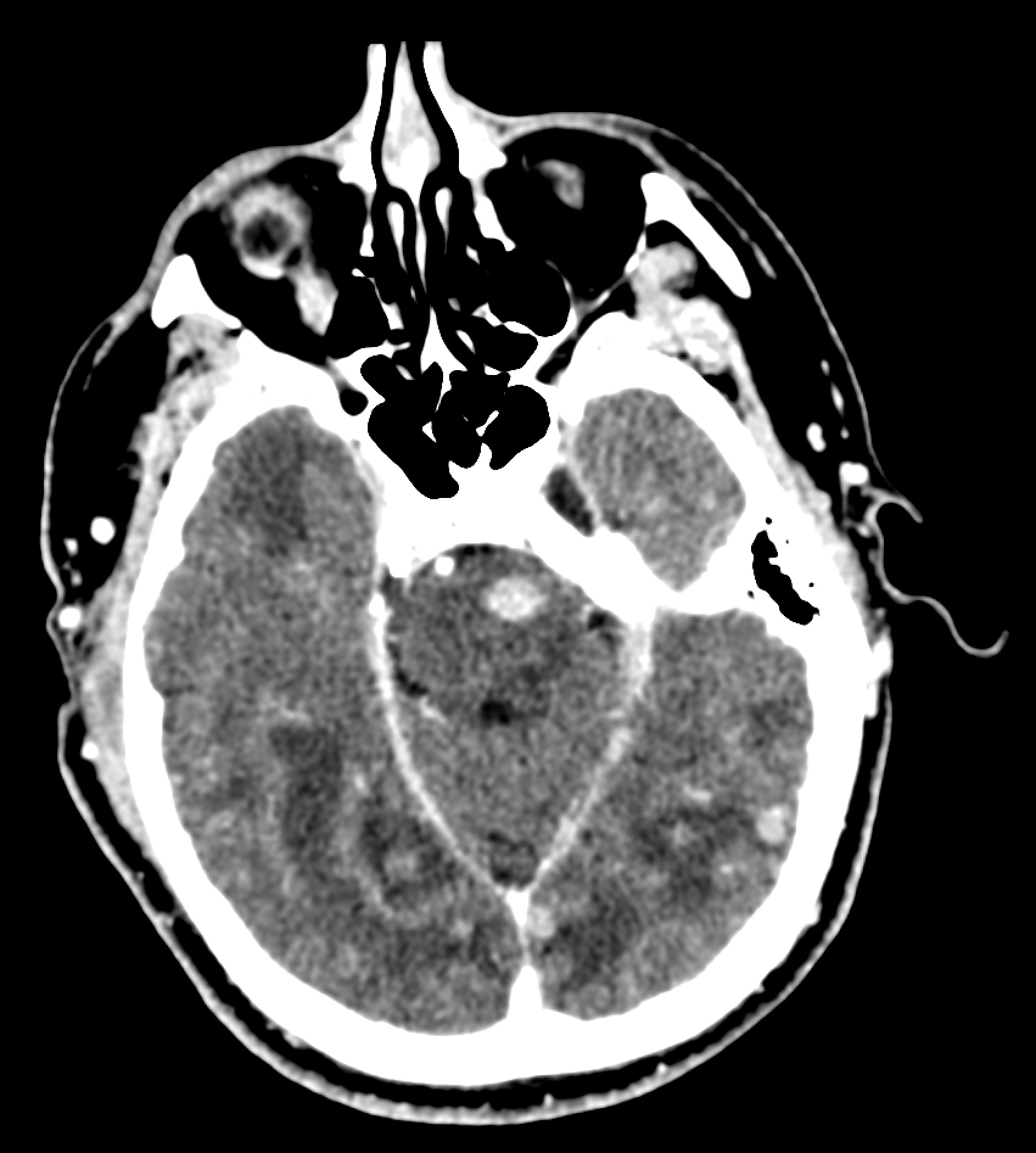
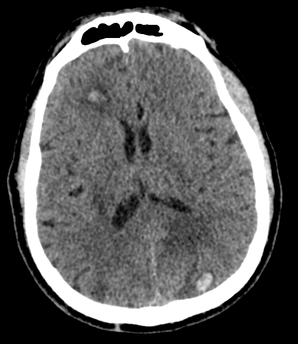
CT brain with and without contrast
The brain lesions are ring-enhancing.
The brain lesions are in a distribution to explain the patient’s clinical symptoms.
The lesions occur in the gray matter.
The lesions are:
On the non-contrast CT, some of the lesions are hemorrhagic.

Watch our video
Second Imaging Study
What is the next imaging study you will order?
Pertinent Imaging Observations
Click on the links below to view images from the study, and assess these key findings as best you can.
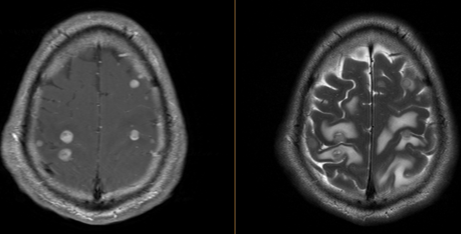
MRI Brain with and without IV contrast
The peritumoral hyperintensity reflects:
Watch our video
What is your Diagnosis now that you have seen the imaging results?
Current Acuity
Initially, you selected and we suggested acuity.
Has your concern for this patient changed?
Assessment and Plan
Please provide your assessment and plan for this patient
Lessons Learned:
- New onset seizures, headaches, and with systemic symptoms in in patients with hx of cancer is concerning for intracranial metastatic disease.
- Metastatic brain tumors are usually ring-enhancing, most often occur in the gray-white junction, and watershed regions. Metastatic melanomas often have intrinsic hemorrhage and therefore appear bright on CT without contrast.
- Surgical resection is the preferred approach for solitary, large tumors, or metastases leading to obstructive hydrocephalus. SRS is preferred when the tumor is small and inaccessible.
- Postoperative stereotactic radiation therapy (SRS) or fractionated radiation therapy to the tumor resection can reduce the risk of local recurrence.
- SRS should be performed when several tumors are present.
- When innumerable tumors are present and systemic therapy is not an option, WBRT is the treatment of choice.
- In patients with a poor prognosis, like those with extracranial disease, the decision for treatment should be performed based on the patient’s preferences.
Socioeconomic Factors: In a patient with known metastatic melanoma, the diagnosis is certain with CT if it shows innumerable hemorrhagic and ring-enhancing metastases. Therefore, the added expense of an MRI is not necessary.
That's the end of the module! Once you've reviewed the video(s), you can click here for another case challenge.
