Retake
C24) Worsening dyspnea and cough in a former shipyard worker
Review the Learning Outcomes, Hx, PE and Labs, and begin the module with your Provisional Diagnosis. Keep hitting "Next" to move through the module.
Learning Outcomes
- Articulate your relationship with the consulting diagnostic radiologists in the evaluation of a patient with shortness of breath.
- Review the DDx considerations in a patient with shortness of breath.
- Identify the spectrum of imaging findings in appropriate modalities for evaluating a patient with shortness of breath.
History
An 80-year-old male presents to clinic with a four-month history of worsening shortness of breath and cough. Prior to retirement, the patient manufactured ships for about 40 years. He denies any associated fevers, chest pain, orthopnea, or leg swelling.
Physical Exam
BP: 128/77, HR: 78, RR: 17, Temp: 99.2 F, O2 saturation 92%.
HEENT: No cervical adenopathy
Cardiac: Normal S1 and S2 heart sounds without murmurs, rubs, or gallops. JVP is 2 cm H2O above sternal angle. No peripheral edema.
Lungs: Fine bibasilar end-inspiratory crackles.
Extremities: There is digital clubbing.
Labs
Pulmonary function testing reveals a low forced expiratory volume (FEV1), low forced vital capacity (FVC), and a FEV1/FVC ratio of 0.82. Diffusing capacity of the lungs for carbon monoxide (DLCO) is also low.
Provisional Diagnosis
Select the Dx you believe is most appropriate
Pneumoconiosis is the most probable provisional diagnosis for the patient who presents with gradually progressive dyspnea, fine bibasilar crackles indicating interstitial lung disease, and a restrictive pattern on pulmonary function testing. The likely etiology is asbestosis, considering the patient's occupational exposure in shipbuilding.
Well done. You were correct
Potential Acuity
What is your assessment of the likely acuity for this patient?
Well done. You were correct
The patient requires routine workup and management as their condition is not immediately life-threatening.
First Imaging Study
What is the first imaging study you will order?
A CXR is an appropriate first imaging modality as it is relatively inexpensive and quickly obtainable.
Well done. You were correct
Pertinent Imaging Observations
Click on the links below to view images from the study, and assess these key findings as best you can.
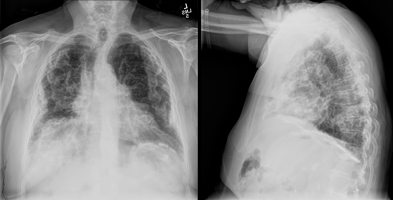
Chest X-ray
What best describes the findings on chest x-ray?
On a chest X-ray, pleural plaques appear as lesions with irregular, thickened, rolled, and nodular margins. They can sometimes be calcified, as in this case. This finding is commonly associated with asbestos exposure and can be an indication of asbestosis.
View the full study if you'd like to take a look yourself.
Second Imaging Study
What is the next imaging study you will order?
A high-resolution CT scan can further characterize lung disease and differentiate it from other non-occupational lung diseases. CT with IV contrast serves no purpose in the setting of suspected ILD. MRI has not been specifically studied for imaging of suspected occupation-associated ILD based on radiography.
Well done. You were correct
Pertinent Imaging Observations
Click on the links below to view images from the study, and assess these key findings as best you can.
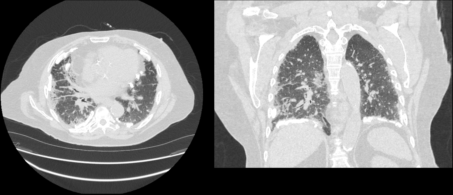
HRCT
Which regions of the lungs are predominantly affected with interstitial fibrosis?
The interstitial fibrosis is predominantly bibasilar, which is a hallmark feature of asbestosis, a lung disease caused by prolonged exposure to asbestos fibers. In contrast, in silicosis, the fibrosis is primarily apical.
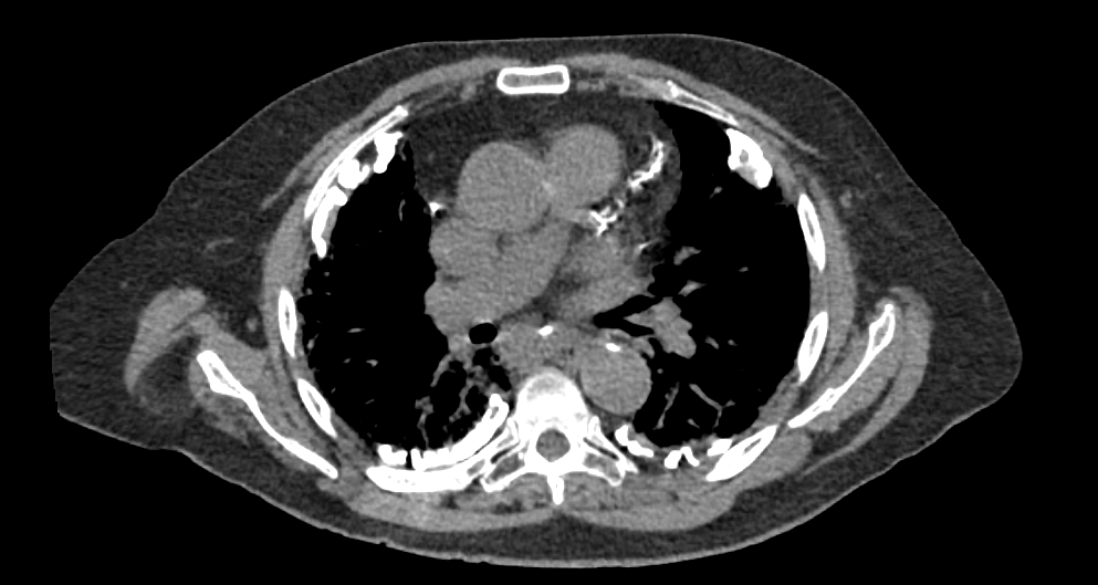
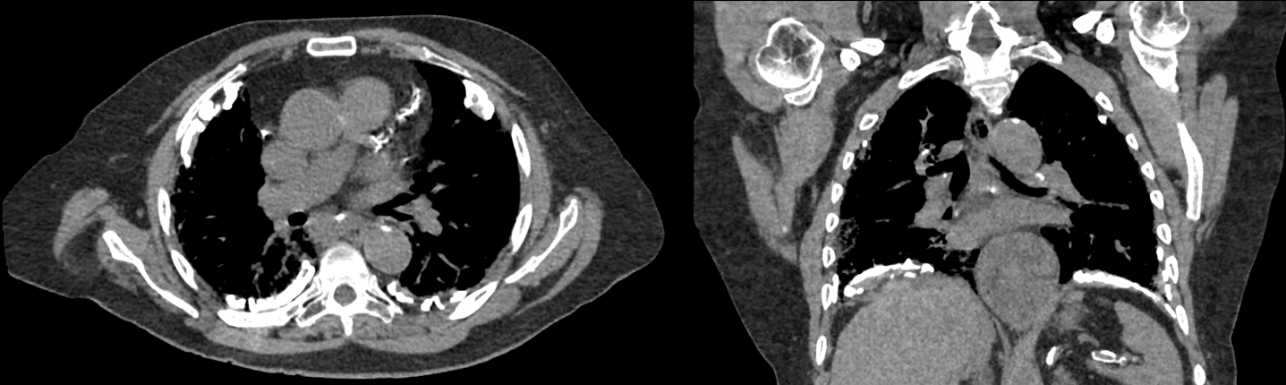
What cardiovascular findings are present?
Extensive coronary artery calcifications are incidentally noted. These calcifications are a marker of atherosclerotic plaque burden, which is a risk factor for cardiovascular disease.
There are other findings.
Diffuse pleural plaques are also present in the supradiaphragmatic region. Pleural plaques are calcified fibrous thickenings of the pleura that often result from exposure to asbestos.
View the full study if you'd like to like a look yourself
Third Imaging Study
What is the next imaging study you will order?
No further imaging is needed as the diagnosis is made with imaging.
What is your Diagnosis now that you have seen the imaging results?
The patient’s presentation, PFTs, and imaging findings are most consistent with asbestosis.
Current Acuity
Initially, you selected and we suggested acuity.
Has your concern for this patient changed?
The patient requires routine management as their condition is not immediately life-threatening.
Assessment and Plan
Please provide your assessment and plan for this patient
This 80-year-old male has presented with shortness of breath over several months, fine inspiratory crackles, and digital clubbing on exam. The high-resolution CT of the chest confirms the diagnosis of asbestosis, with evidence of bilateral pleural plaques and basilar interstitial fibrosis. As there is no curative treatment for asbestosis, management will focus on preventing progression and reducing risk factors for lung cancer. Appropriate measures will include oxygen therapy, avoidance of exposure, smoking cessation, and immunization against influenza virus and pneumococcal pneumonia.
Lessons Learned:
- Asbestosis is a type of pneumoconiosis caused by prolonged exposure to asbestos in industries such as shipbuilding, mining, insulation, and pipe work.
- Asbestos exposure can also cause bronchogenic carcinoma and mesothelioma.
- Symptoms typically appear after a latency period of 20+ years and may include progressive dyspnea, fine basilar crackles, and clubbing.
- Chest X-rays can show bilateral pleural plaques and reticular opacities with basilar predominance. A high-resolution CT scan can further characterize the diagnosis and differentiate it from other non-occupational lung diseases.
- Pulmonary function testing will show a restrictive pattern of disease due to interstitial lung disease caused by asbestosis.
- Late complications can include pulmonary hypertension secondary to hypoxia-induced vasoconstriction, which can eventually lead to cor pulmonale.
- Management of asbestosis focuses on reducing risk factors and slowing disease progression, as there are currently no effective treatments.
That's the end of the module! Once you've reviewed the video(s), you can click here for another case challenge.
Next

