C4) Acute respiratory distress
Review the Learning Outcomes, Hx, PE and Labs, and begin the module with your Provisional Diagnosis. Keep hitting "Next" to move through the module.
Learning Outcomes
- Articulate your relationship with the consulting diagnostic radiologists in the evaluation of a patient with fever and cough.
- Review the DDx considerations in fever and cough.
- Identify the spectrum of imaging findings in appropriate modalities for evaluating patients with fever and cough.
History
Physical Exam
Labs
Provisional Diagnosis
Potential Acuity
What is your assessment of the likely acuity for this patient?
First Imaging Study
What is the first imaging study you will order?
Pertinent Imaging Observations
Click on the links below to view images from the study, and assess these key findings as best you can.
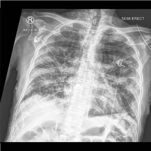
Chest X-ray
What best describes the findings on the Chest X-ray?

Watch our video
Second Imaging Study
What is the next imaging study you will order?
Pertinent Imaging Observations
Click on the links below to view images from the study, and assess these key findings as best you can.
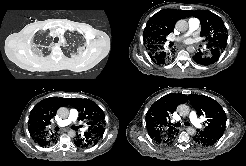
Chest CT
What best describes the findings on the Chest CT?
Watch our video
Third Imaging Study
What is the next imaging study you will order?
What is your Diagnosis now that you have seen the imaging results?
Current Acuity
Initially, you selected and we suggested acuity.
Has your concern for this patient changed?
Assessment and Plan
Please provide your assessment and plan for this patient
Lessons Learned: The hypercoagulable state secondary to COVID-19 infection can lead to a pulmonary embolism. COVID-19 pneumonia can appear on chest x-ray as multifocal pneumonia with numerous airspace consolidations. CT findings characteristic for COVID-19 pneumonia include diffuse ground glass opacities with basilar and peripheral predominance, air space consolidations, bronchovascular thickening, and traction bronchiectasis.
Socioeconomic Factors: Patients with high clinical suspicion for COVID-19 infection should be immediately tested and placed under droplet precautions prior to imaging, if possible. Portable imaging for the initial chest x-ray is preferable to minimize infection spread.
That's the end of the module! Once you've reviewed the video(s), you can click here for another case challenge.
Contributors:
Kevin Pierre, MD - Editor
Robbie Slater, MD - Supervising Editor
Bayar Batmunh, MS - Coordinator
