Retake
A6) RUQ abdominal pain and weight loss with a history of breast cancer
Review the Learning Outcomes, Hx, PE and Labs, and begin the module with your Provisional Diagnosis. Keep hitting "Next" to move through the module.
Learning Outcomes
- Articulate your relationship with the consulting diagnostic radiologists in the evaluation of a patient with abdominal pain.
- Identify the spectrum of imaging findings in appropriate modalities for evaluating a patient with abdominal pain.
History
A 75-year-old female with a history of breast cancer treated with bilateral mastectomy, chemotherapy, and radiation therapy presents with a 2-month history of progressively worsening dull, achy right upper quadrant pain accompanied by an unintentional 18 lb weight loss.
Physical Exam
BP: 106/65, HR 98, RR 18, Temp 98.6 deg F, O2 saturation 100% on room air.
General: Awake, alert, and cachectic.
Skin: Jaundiced and pale.
Eyes: Scleral icterus present.
Abdomen: Tenderness in the right upper quadrant.
Labs
CBC:
Hb: 9.5 gm/dL (Normal Range: 13.5 – 17.5 gm/dL),
MCV: 80.1 fL (Normal Range: 80 – 100 fL),
WBC: 7,500 /µL (Normal Range: 4,500 - 11,000 /µL).
LFTs:
Total Bilirubin: 5.3 mg/dL (Normal Range: 0.1 - 1.0 mg/dL),
Direct Bilirubin: 2.8 mg/dL (Normal Range: 0.0 – 0.3 mg/dL),
ALT: 78 U/L (Normal Range: 10-40 U/L),
AST: 306 U/L (Normal Range: 12-38 U/L),
ALP: 640 U/L (Normal Range: 25-100 U/L).
ESR:
75 mm/h (Normal Range: 0 – 20 mm/h).
Provisional Diagnosis
Select the Dx you believe is most appropriate
Given the patient's history of breast cancer, her presentation with non-specific right upper quadrant abdominal pain and significant unintentional weight loss over several months, coupled with physical exam findings of jaundice, ascites, and hepatomegaly, and the abnormal pattern of LFTs, it is highly suggestive of potential liver metastasis from the primary breast cancer.
Well done. You were correct
Potential Acuity
What is your assessment of the likely acuity for this patient?
Well done. You were correct
The patient requires routine, but expedited workup.
First Imaging Study
What is the first imaging study you will order?
Ultrasound of the right upper quadrant of the abdomen is an appropriate initial choice to evaluate the liver parenchyma.
Well done. You were correct
Pertinent Imaging Observations
Click on the links below to view images from the study, and assess these key findings as best you can.
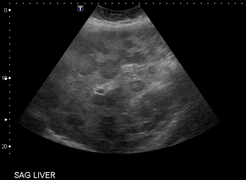
Abdominal ultrasound
What best describes the findings on the abdominal ultrasound?
There are multiple liver lesions, which likely represent metastases in this patient with a history of breast cancer.
View the full study if you'd like to take a look yourself.
Second Imaging Study
What is the next imaging study you will order?
A contrast-enhanced CT can help identify and differentiate between metastases and benign lesions in patients with a history of primary malignancy. In this case, a CT chest was also obtained to further evaluate for suspected metastatic breast cancer.
Well done. You were correct
Pertinent Imaging Observations
Click on the links below to view images from the study, and assess these key findings as best you can.
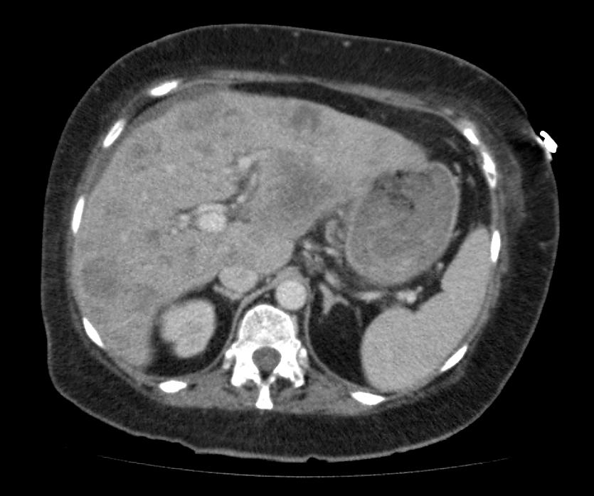
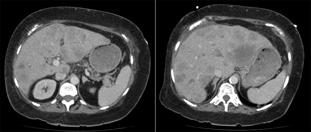
CT chest, abdomen, and pelvis with contrast
The liver lesions are:
The liver lesions are hypoattenuating to the remainder of the liver parenchyma and demonstrate peripheral enhancement. These findings are highly indicative of liver metastases.
There is evidence of metastases to other organs.
Yes, there are bilateral adrenal nodules that likely represent metastatic disease.
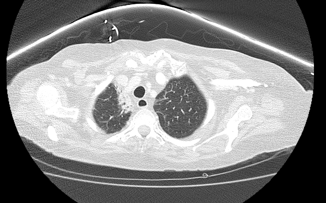
The upper lung fibrosis is likely secondary to:
The upper lobe changes are likely secondary to radiation therapy for her breast cancer, as the patient has no occupational exposure.
View the full study if you'd like to like a look yourself
Third Imaging Study
What is the next imaging study you will order?
A PET may be ordered to detect and evaluate the extent of metastatic breast cancer beyond the liver. Additionally, a PET scan serves as a baseline for monitoring the patient's response to treatment over time. These images are not shown here.
What is your Diagnosis now that you have seen the imaging results?
The patient’s history and imaging suggest breast cancer metastases to the liver and adrenal glands.
Current Acuity
Initially, you selected and we suggested acuity.
Has your concern for this patient changed?
The patient requires routine, but expedited workup and management.
Assessment and Plan
Please provide your assessment and plan for this patient
This is a 72-year-old female patient with a history of breast cancer who has undergone bilateral mastectomy, chemotherapy, and radiation therapy. She is currently presenting with vague right upper quadrant pain persisting for 2 months, unintentional weight loss of 18 lbs, and elevated liver function tests (LFTs). Abdominal ultrasound and subsequent CT scan of the chest, abdomen, and pelvis with IV contrast reveal multiple hypodense lesions in the liver and bilateral adrenal nodules, raising concerns for metastatic disease. Considering the patient’s history, these lesions likely represent metastases from breast cancer. To confirm the diagnosis, arranging a biopsy is recommended.
The patient should be referred to a multidisciplinary team comprising medical, surgical, and radiation oncologists, as well as supportive care specialists. Treatment options will be tailored based on tumor characteristics, molecular profiling, and the patient's previous treatment history. Systemic chemotherapy or immunotherapy may be considered as appropriate.
Early involvement of palliative care services is crucial to address symptom control, provide psychosocial support, and assist the patient in making informed decisions about her care. Encouraging active participation in treatment decisions, the patient should be provided with necessary resources and education to cope with her diagnosis and navigate the treatment journey effectively.
Lessons Learned:
- Common primary tumor sites associated with metastatic liver disease include the colon, stomach, pancreas, lung, and breast.
- Abdominal ultrasound serves as an appropriate initial assessment for detecting potential liver metastasis.
- Ultrasound classically reveals multiple hypoechoic lesions with hypoechoic halos within the liver parenchyma.
- Following an initial assessment, a CT scan of the abdomen with intravenous contrast is recommended to provide detailed imaging of the liver and surrounding structures.
- CT commonly reveals multiple hypodense, ring-enhancing lesions. Solitary metastases are relatively uncommon.
- Lung fibrosis can be a potential consequence of radiation therapy.
Socioeconomic Factors: African American women experience higher breast cancer mortality rates compared to women of other ethnic backgrounds, which may be attributed to lower rates of screening, delayed diagnosis, and inadequate healthcare accessibility.
That's the end of the module! Once you've reviewed the video(s), you can click here for another case challenge.
Next

