Retake
A3) Abnormal liver function tests
Review the Learning Outcomes, Hx, PE and Labs, and begin the module with your Provisional Diagnosis. Keep hitting "Next" to move through the module.
Learning Outcomes
- Articulate your relationship with the consulting diagnostic radiologists in the evaluation of a patient with an incidental finding.
- Identify the spectrum of imaging findings in appropriate modalities for evaluating a patient with an incidental finding.
History
A 40-year-old with hyperlipidemia and coronary artery disease was started on statins by her PCP. Follow-up routine liver function tests were persistently abnormal, even after discontinuing the statins. She denies any symptoms. She also denies a history of cirrhosis or malignancy.
Physical Exam
BP: 128/76, HR 68, RR 15, Temp 37ºC, O2 saturation 99% BMI 27
Abdominal: No abdominal tenderness.
Labs
BMI: 24 (nl: 18.5 to 24.9 kg/m²).
ALT: 45 U/L (nl: 5-40 U/L),
AST: 38 U/L (nl: 9-32 U/L),
ALP: 128 U/L (nl: 44-147 U/L).
Serology negative for hepatitis A, B, and C.
AFP within normal limits.
Provisional Diagnosis
Select the Dx you believe is most appropriate
The patient’s elevated LFTs is most likely secondary to a benign liver mass considering the lack of signs, symptoms, or physical exam findings.
Well done. You were correct
Potential Acuity
What is your assessment of the likely acuity for this patient?
Well done. You were correct
The patient requires routine workup as their condition is not immediately life-threatening.
First Imaging Study
What is the first imaging study you will order?
An abdominal ultrasound is an appropriate and cost-effective initial imaging modality that avoids exposure to radiation.
Well done. You were correct
Pertinent Imaging Observations
Click on the links below to view images from the study, and assess these key findings as best you can.
Abdominal US with doppler
There is evidence of acute cholecystitis.
There is no gallbladder wall thickening or pericholecystitic fluid to suggest acute cholecystitis. Furthermore, there are no gallstones.
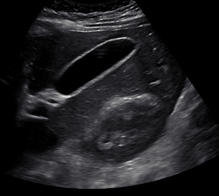
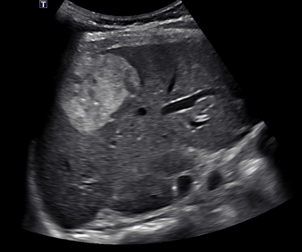
There is a liver lesion.
There is a well circumscribed, homogenously lesion in the liver.
The liver lesion is:
The liver lesion is echogenic, or hyperechoic.
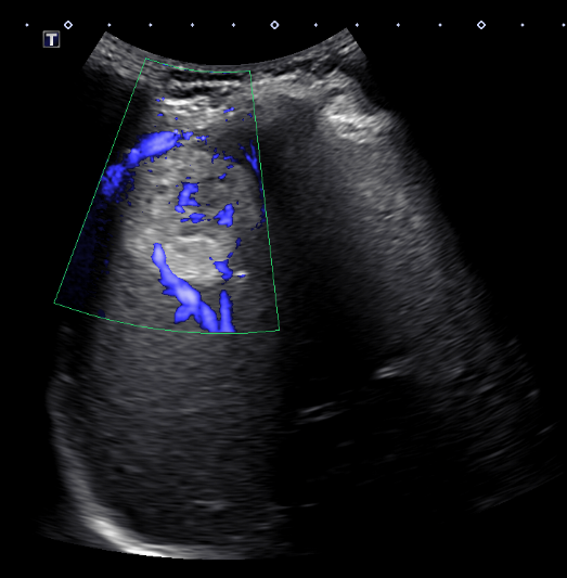
There is blood flow through the liver lesion.
Color doppler demonstrates blood flow through the liver lesion.
View the full study if you'd like to take a look yourself.
Second Imaging Study
What is the next imaging study you will order?
No further imaging is needed as the patient does not have any history of extrahepatic malignancy or cirrhosis. However, we show a CT to demonstrate the characteristic studies in case a CT had been ordered.
Well done. You were correct
Pertinent Imaging Observations
Click on the links below to view images from the study, and assess these key findings as best you can.
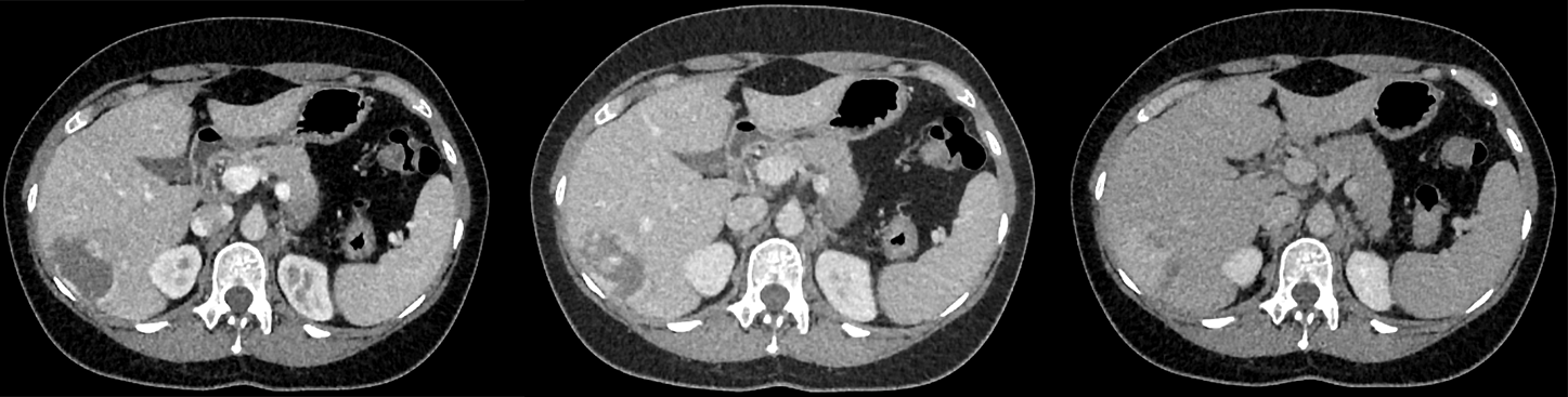
CT abdomen with IV contrast
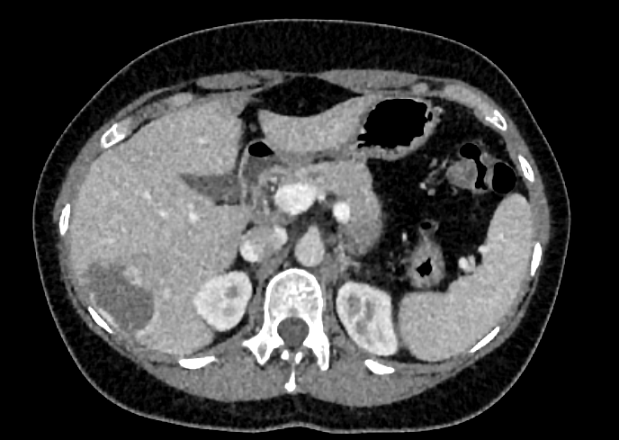
The liver lesion is:
The lesion is darker than the remainder of the liver parenchyma, and is therefore hypoattenuating.
When comparing the arterial, venous, and delayed phases, the liver lesion demonstrates:
The lesion demonstrates peripheral, discontinuous, nodular enhancement on the arterial phase. The lesion then becomes progressively hyperintense during the venous then delayed phase, demonstrating central fill in. On the other hand, washout, which is characteristic of hepatocellular carcinoma, refers to progressive clearance of contrast material from the center of the lesion.
View the full study if you'd like to like a look yourself
Third Imaging Study
What is the next imaging study you will order?
No further imaging is required as the diagnosis is strongly suspected with the CT scan.
What is your Diagnosis now that you have seen the imaging results?
Liver hemangiomas are benign liver masses often diagnosed incidentally.
Current Acuity
Initially, you selected and we suggested acuity.
Has your concern for this patient changed?
The patient requires routine management as their condition is not immediately life-threatening
Assessment and Plan
Please provide your assessment and plan for this patient
This is a 40-year-old female with an incidentally discovered benign liver hemangioma. No further imaging is required as the liver lesion is less than 5 cm. The patient should continue to undergo routine liver function tests with their primary care provider. They should also undergo a follow-up for their incidentally noted lung nodule. Given that this is asymptomatic, treatment is not necessary.
Lessons Learned:
- Hepatic hemangiomas are a type of vascular malformation that occurs within the liver parenchyma.
- Hemangiomas are the most common type of benign liver lesion, and they are typically diagnosed incidentally during imaging studies.
- Although hemangiomas occur in both sexes, they are more common in women.
- Hemangiomas demonstrate blood flow and peripheral feeding vessels on Doppler ultrasound and show a characteristic "filling in" of contrast on contrast-enhanced CT.
- Management of hepatic hemangiomas typically involves observation, with intervention reserved for symptomatic or enlarging lesions.
- Although complications of hepatic hemangiomas are rare, giant hemangiomas can cause Kasabach-Merritt syndrome, or hemolytic anemia, thrombocytopenia, and hypofibrinogenemia.
That's the end of the module! Once you've reviewed the video(s), you can click here for another case challenge.
Next

