R8) Patient presenting for breast cancer screening
Review the Learning Outcomes, Hx, PE and Labs, and begin the module with your Provisional Diagnosis. Keep hitting "Next" to move through the module.
Learning Outcomes
- Articulate your relationship with the consulting diagnostic radiologists in the evaluation of a patient with a breast mass.
- Review the DDx considerations in a breast mass.
- Identify the spectrum of imaging findings in appropriate modalities for evaluating patients with a breast mass.
History
Physical Exam
Labs
Provisional Diagnosis
Potential Acuity
What is your assessment of the likely acuity for this patient?
First Imaging Study
What is the first imaging study you will order?
Pertinent Imaging Observations
Click on the links below to view images from the study, and assess these key findings as best you can.
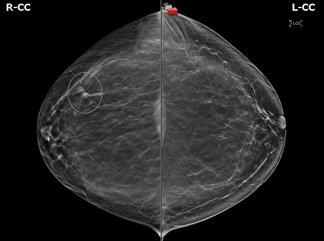
Mammogram
There is a mass present.
The mass is located in which quadrant of the breast?
The mass is:

Watch our video
Second Imaging Study
What is the next imaging study you will order?
Pertinent Imaging Observations
Click on the links below to view images from the study, and assess these key findings as best you can.
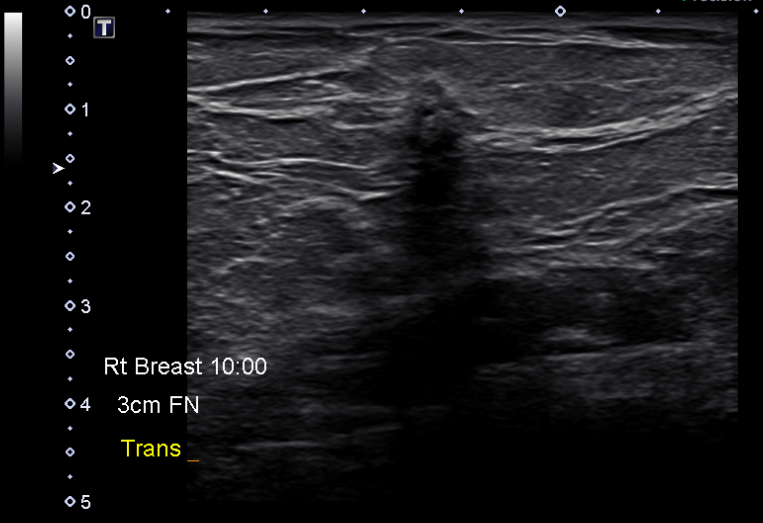
Breast ultrasound
The features of the mass suggest it is:
Watch our video
Third Imaging Study
What is the next imaging study you will order?
What is your Diagnosis now that you have seen the imaging results?
Current Acuity
Initially, you selected and we suggested acuity.
Has your concern for this patient changed?
Assessment and Plan
Please provide your assessment and plan for this patient
Lessons Learned:
- As of 2023, The United States Preventive Services Task Force (USPSTF) recommends regular screening mammography for women between the ages of 40 and 74, with a frequency of once every two years.
- A palpable mass is often the primary symptom of breast cancer. Furthermore, palpable cancers tend to exhibit aggressive behavior and have a less favorable prognosis compared to cancers detected through regular screening.
- For women over 40, a diagnostic mammogram is the recommended initial imaging technique for evaluating a palpable breast mass. In cases where there is uncertainty regarding the correlation between the mammographic finding and the palpable lesion, a targeted ultrasound can be used as a secondary study.
- Mammographic features that may suggest malignancy include irregular masses, spiculation, and the presence of microcalcifications.
- Ultrasound features that may suggest malignancy include ill-defined, hypoechoic masses, hyperechoic angular margins, posterior acoustic shadowing, posterior enhancement, a branched or spiculated pattern, being “taller than broader,” and the presence of microcalcifications.
Socioeconomic Factors: Women with higher socioeconomic status show significantly higher breast cancer incidence, which may be explained by reproductive factors, mammography screening, hormone replacement therapy and lifestyle factors.
That's the end of the module! Once you've reviewed the video(s), you can click here for another case challenge.
