Retake
R12) Vaginal bleeding in a patient with an enlarged uterus
Review the Learning Outcomes, Hx, PE and Labs, and begin the module with your Provisional Diagnosis. Keep hitting "Next" to move through the module.
Learning Outcomes
- Articulate your relationship with the consulting diagnostic radiologists in the evaluation of patient with vaginal bleeding.
- Review the DDx considerations in a patient with vaginal bleeding.
- Identify the spectrum of imaging findings in appropriate modalities for evaluating a patient with vaginal bleeding.
History
A 24-year-old female presents to the ED with an episode of light vaginal bleeding that occurred following intercourse and resolved within 30 minutes. She denies any associated abdominal pain, cramping, or other distressing symptoms. Her last menstrual period was approximately 11 weeks ago. She acknowledges the possibility of being pregnant but has not confirmed it with a pregnancy test. Her last medical check-up was 3 years ago. She denies any history of sexually transmitted infections, urinary tract infections, or abnormal Pap smear results. She also denies any symptoms suggestive of pregnancy such as contractions or perception of fetal movement. Today's episode was her first occurrence of vaginal bleeding since her last menstrual period.
Physical Exam
Vitals: Blood pressure is 125/80 mmHg, heart rate is 88 beats per minute, respiratory rate is 12 breaths per minute, temperature is 99.8 F, and O2 saturation is 98% on room air.
• External: The external genitalia appear normal without any lesions or discharge.
• Speculum examination: A small amount of dark red blood is pooled in the posterior fornix. The cervix appears normal and is closed.
• Bimanual examination: No adnexal, uterine, or cervical motion tenderness noted. The uterus is enlarged with fundal height measuring 11 cm, which is consistent with an estimated gestational age of approximately 11 weeks.
Labs
Beta-HCG: 50,000 IU/L
Provisional Diagnosis
Select the Dx you believe is most appropriate
The patient is most likely pregnant considering the elevated bHCG, LMP about 11 weeks ago, and an enlarged uterus. Small amounts of vaginal bleeding may occur throughout the course of a normal pregnancy but warrant evaluation.
Well done. You were correct
Potential Acuity
What is your assessment of the likely acuity for this patient?
Well done. You were correct
The patient requires routine workup and management. They are hemodynamically stable and no longer experiencing vaginal bleeding.
First Imaging Study
What is the first imaging study you will order?
Transvaginal ultrasound (TVUS) is a highly sensitive modality for confirming pregnancy, estimating gestational age, and evaluating for any potential complications. Furthermore, it can rule out other causes of bleeding such as fibroids or placenta previa.
Well done. You were correct
Pertinent Imaging Observations
Click on the links below to view images from the study, and assess these key findings as best you can.
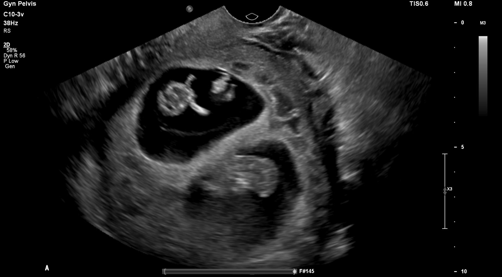
Transvaginal ultrasound
What best describes the findings on the transvaginal ultrasound?
There are two distinct gestational sacs, each with a fetus. The membrane separating the twins appears thickened, displaying the characteristic "lambda" or "twin peak" sign. This sign indicates the presence of two separate placentas or chorions, which is characteristic of a dichorionic-diamniotic twin pregnancy.
There is another finding.
The hypoechoic area adjacent to the cervical os represents a subchorionic perigestational hemorrhage.
View the full study if you'd like to take a look yourself.
Second Imaging Study
What is the next imaging study you will order?
A formal anatomy ultrasound should be performed between 18 and 22 weeks of gestation to assess fetal anatomy, ensure proper development, and evaluate the location of the placenta and the amount of amniotic fluid.
Well done. You were correct
What is your Diagnosis now that you have seen the imaging results?
A dichorionic-diamniotic twin pregnancy is indicated by two separate placentas, a thick intertwin membrane, and the presence of the 'lambda' sign on ultrasound. In contrast, a monochorionic-monoamniotic pregnancy, where twins share a placenta and amniotic sac, shows no intertwin membrane. Therefore, based on these ultrasound features, the likely diagnosis is a dichorionic-diamniotic twin pregnancy.
Current Acuity
Initially, you selected and we suggested acuity.
Has your concern for this patient changed?
The patient requires routine workup and management.
Assessment and Plan
Please provide your assessment and plan for this patient
This patient, a 24-year-old G1P0 female, presents with a single episode of light vaginal bleeding following intercourse, which has since resolved. Her beta-HCG levels, last menstrual period approximately 11 weeks ago, and physical examination findings were all consistent with a diagnosis of pregnancy. A transvaginal ultrasound confirmed a dichorionic-diamniotic twin pregnancy, and also identified a small subchorionic perigestational hemorrhage near the cervical os, which is likely the cause of her bleeding. This is a common cause of first-trimester bleeding and given the self-limited bleeding and stable status, she can be discharged with a follow-up ultrasound planned. We should also establish prenatal care for the patient, including prenatal vitamins and first-trimester labs.
Lessons Learned:
- There is an increasing incidence of multifetal pregnancies due to factors such as increased maternal age at conception and increasing use of assisted reproductive technology.
- Twin pregnancies can be classified into four types: dichorionic-diamniotic, monochorionic-diamniotic, monochorionic-monoamniotic, and conjoined twins, each with their own unique potential complications, monitoring strategies, and delivery planning.
- Perigestational hemorrhage, with varying presentations and locations, has a 90% successful outcome in cases of minor bleeding accompanied by reassuring fetal monitoring; however, twin pregnancies pose additional risks for this type of hemorrhage.
- Subchorionic hemorrhage, most prevalent during 9-20 weeks of gestational age, is the leading cause of bleeding in the first trimester. If bleeding is limited and both fetal monitoring and maternal vital signs are stable, the patient can be safely discharged with a follow-up ultrasound scheduled.
That's the end of the module! Once you've reviewed the video(s), you can click here for another case challenge.
Next

