R10) Enlarging breast mass in a 22-year-old female
Review the Learning Outcomes, Hx, PE and Labs, and begin the module with your Provisional Diagnosis. Keep hitting "Next" to move through the module.
Learning Outcomes
- Articulate your relationship with the consulting diagnostic radiologists in the evaluation of a patient with a breast mass.
- Review the DDx considerations in a breast mass.
- Identify the spectrum of imaging findings in appropriate modalities for evaluating patients with a breast mass.
History
Physical Exam
Labs
Provisional Diagnosis
Potential Acuity
What is your assessment of the likely acuity for this patient?
First Imaging Study
What is the first imaging study you will order?
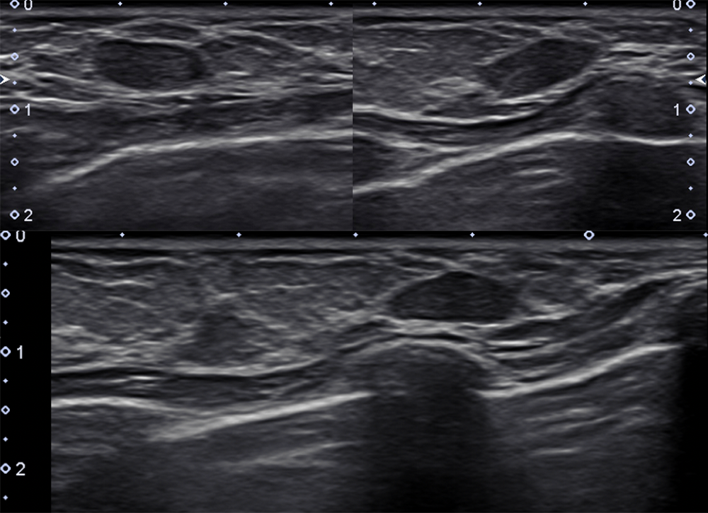
Pertinent Imaging Observations
Click on the links below to view images from the study, and assess these key findings as best you can.

Watch our video
Second Imaging Study
What is the next imaging study you will order?
What is your Diagnosis now that you have seen the imaging results?
Current Acuity
Initially, you selected and we suggested acuity.
Has your concern for this patient changed?
Assessment and Plan
Please provide your assessment and plan for this patient
Lessons Learned:
- Fibroadenomas are estrogen-responsive tumors. As such, they are most likely to occur in patients who are of reproductive age, pregnant, and lactating. They enlarge in the pre-ovulation phase of the menstrual cycle.
- Most fibroadenomas occur in the upper outer quadrant and are well-circumscribed, soft, and mobile.
- The best diagnostic modality in a patient younger than 40 is an ultrasound. Younger patients are more likely to have dense breasts which can obscure lesions on mammography.
- Ultrasound findings reassuring that the mass is a benign fibroadenoma include homogeneous echotexture, well-defined margins, and sometimes a “pseudocapsule.”
- The diagnosis of a benign fibroadenomas can be confirmed with a core-needle biopsy or followed with serial short-term (3-6 month) imaging and breast examination. A complex fibroadenoma that continues to grow increases risk of breast cancer and warrants an ultrasound-guided biopsy. Fibroadenomas greater than 2.5cm without previous imaging should undergo biopsy.
That's the end of the module! Once you've reviewed the video(s), you can click here for another case challenge.
