Retake
P20) Developmental delay and ataxia
Review the Learning Outcomes, Hx, PE and Labs, and begin the module with your Provisional Diagnosis. Keep hitting "Next" to move through the module.
Learning Outcomes
- Articulate your relationship with the consulting diagnostic radiologists in the evaluation of a pediatric patient with ataxia.
- Review the DDx considerations in a pediatric patient with ataxia.
- Identify the spectrum of imaging findings in appropriate modalities for evaluating a pediatric patient with ataxia.
History
A one-year-old male infant is brought into the pediatric clinic by his mother due to concerns about delayed developmental milestones. She reports her son's struggle with balance and coordination skills, such as rolling over and sitting up, which his peers have achieved by this age. Additionally, she has observed increased irritability and an unusual increase in her son's head size over the past few months. The child was born at full term; however, the mother did not receive routine prenatal care due to socioeconomic limitations.
Physical Exam
Vitals: BP: 90/52, HR: 110 bpm, RR: 30/min, Temp: 99 F, O2 saturation: 95%.
HEENT: The child's head circumference is above the 97th percentile for his age.
Neurological Examination: Hypotonia and decreased motor coordination.
Labs
None
Provisional Diagnosis
Select the Dx you believe is most appropriate
The most probably diagnosis appears to be a posterior fossa malformation, possibly Dandy-Walker malformation, which is characterized by the findings of macrocephaly and developmental delay. While spina bifida and Chiari malformation could present with similar symptoms, the absence of spinal abnormalities and other pertinent physical exam findings make these diagnoses less likely. Holoprosencephaly is usually associated with severe craniofacial abnormalities, which are not present in this case.
Well done. You were correct
Potential Acuity
What is your assessment of the likely acuity for this patient?
Well done. You were correct
The patient will require routine workup and management as their condition is not immediately life-threatening.
First Imaging Study
What is the first imaging study you will order?
MRI is the preferred initial imaging modality in a child with ataxia due to its higher sensitivity in detecting structural developmental disorders, infectious and postinfectious disorders, demyelinating conditions, and brain tumors, which are common causes of acute cerebellar ataxia in children.
Well done. You were correct
Pertinent Imaging Observations
Click on the links below to view images from the study, and assess these key findings as best you can.
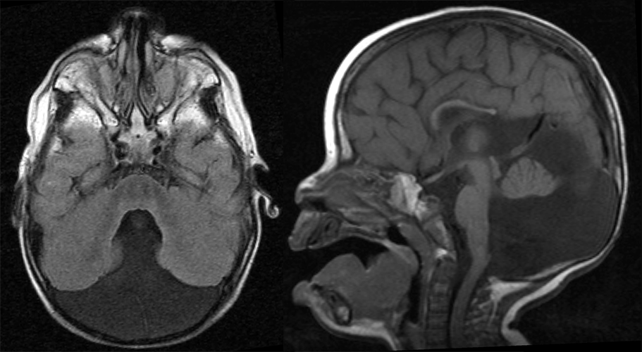
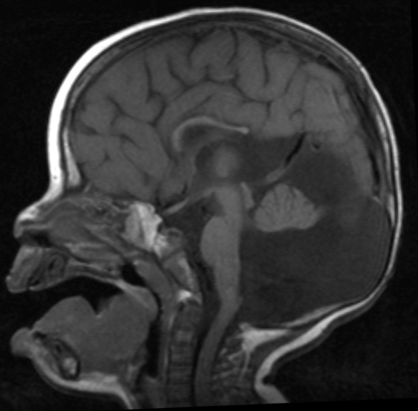
Brain MRI
There is an enlarged posterior fossa.
There is cystic dilatation of the fourth ventricle with anterior displacement of the cerebellum leading to enlargement of the posterior fossa. This finding, combined with the hypoplasia of the vermis, is suggestive of Dandy Walker malformation.
There are other abnormalities.
The corpus callosum is hypoplastic. This finding may be seen in patients with Dandy Walker malformations.
View the full study if you'd like to take a look yourself.
Second Imaging Study
What is the next imaging study you will order?
No further imaging is required as the diagnosis is confirmed with the MRI.
Well done. You were correct
What is your Diagnosis now that you have seen the imaging results?
The presence of ataxia in a 1 year old with imaging findings of cystic dilation of the posterior fossa and hypoplasia of the vermis is indicative of a Dandy-Walker malformation.
Current Acuity
Initially, you selected and we suggested acuity.
Has your concern for this patient changed?
The patient will require routine workup and management as their condition is not immediately life-threatening.
Assessment and Plan
Please provide your assessment and plan for this patient
The patient is a 1-year-old male who presents with ataxia, delayed developmental milestones, and increased head size. On physical examination, the patient has macrocephaly, hypotonia, and decreased motor coordination. The MRI demonstrates findings of a Dandy-Walker malformation with cystic dilation of the posterior fossa and hypoplasia of the vermis. A pediatric neurologist and a pediatric neurosurgeon should be consulted to assess the need for intervention such as ventriculoperitoneal shunting, considering the ventricular dilation. The patient’s parents should also be referred to a genetic counselor. The patient should also be scheduled for recommended childhood vaccinations and well-child visits.
Lessons Learned:
- Dandy-Walker malformation is a congenital brain malformation characterized by an enlarged fourth ventricle and incomplete formation or absence of the cerebellar vermis. This condition often results in a cystic enlargement of the fourth ventricle, which accumulates cerebrospinal fluid (CSF), causing an expansion of the posterior fossa and obstructive/noncommunicative hydrocephalus.
- Patients with Dandy-Walker malformation may develop hydrocephalus.
- Manifestations of Dandy-Walker malformation can vary, but often include macrocephaly, developmental delays, changes in behavior, and in some cases, decreased levels of consciousness.
- Prenatal diagnosis is possible using ultrasound. Specific markers include the cystic dilation of the fourth ventricle, presence of a persistent Blake's pouch, and an abnormally large posterior fossa.
- For older infants and adults, MRI is the preferred imaging modality. Characteristic findings are cystic dilation of the posterior fossa and vermic hypoplasia.
- Treatment often involves placement of a cerebral shunt. This device helps to drain the excess CSF from the brain into the peritoneal cavity, alleviating the symptoms caused by increased intracranial pressure.
Socioeconomic Factors: Dandy-Walker malformations are typically identified during the prenatal period, often around the 18th week of gestation, using ultrasound imaging. However, socioeconomic disparities can impact the accessibility to and utilization of such prenatal care. Individuals from lower socioeconomic strata may face barriers in obtaining routine prenatal screenings, which could potentially lead to a delayed diagnosis.
That's the end of the module! Once you've reviewed the video(s), you can click here for another case challenge.
Next

