Retake
P19) Worsening nocturnal knee pain
Review the Learning Outcomes, Hx, PE and Labs, and begin the module with your Provisional Diagnosis. Keep hitting "Next" to move through the module.
Learning Outcomes
- Articulate your relationship with the consulting diagnostic radiologists in the evaluation of a pediatric patient with extremity pain.
- Review the DDx considerations in a pediatric patient with extremity pain.
- Identify the spectrum of imaging findings in appropriate modalities for evaluating a pediatric patient with extremity pain.
History
A 16-year-old male has been experiencing worsening pain in his right knee, which is particularly severe at night. Over the past five months, this nocturnal pain has been significant enough to awaken him from sleep. The pain is also causing him to limp, and over-the-counter analgesics, which initially provided relief, are no longer effective. Additionally, he has reported unexplained episodes of fever and weight loss in the past.
Physical Exam
BP: 120/83, HR 80/min, RR 13/min, Temp 97.8 F, O2 saturation 99%. Tenderness to palpation with swelling and warmth over the right fever. Neurovascular examination of the right leg is intact.
Labs
• Hemoglobin: 12.2 g/dL (Reference range: 13.5-17.5 g/dL)
• White Blood Cell Count (WBC): 7.2 x10^9/L (Reference range: 4.5-11.0 x10^9/L)
• Erythrocyte Sedimentation Rate (ESR): 50 mm/hr (Reference range: 0-15 mm/hr)
• C-Reactive Protein (CRP): 25 mg/L (Reference range: <10 mg/L)
• Alkaline Phosphatase (ALP): 350 U/L (Reference range: 44-147 U/L)
• Lactate Dehydrogenase (LDH): 450 U/L (Reference range: 105-333 U/L)
Provisional Diagnosis
Select the Dx you believe is most appropriate
The most appropriate diagnosis for this patient is a primary bone tumor. Osteosarcoma typically presents in adolescents with persistent, progressively worsening pain, often severe enough to disrupt sleep. It may also cause systemic symptoms like fever and weight loss. Moreover, the elevated ALP and LDH in the lab results further support the diagnosis.
Well done. You were correct
Potential Acuity
What is your assessment of the likely acuity for this patient?
Well done. You were correct
The patient requires routine, but expedited workup.
First Imaging Study
What is the first imaging study you will order?
Radiographs are an appropriate initial imaging modality as they identify destructive sclerotic lesions and periosteal reactions of the bone. They can offer information regarding the tumor's location, size, shape, margins, and biological activity.
Well done. You were correct
Pertinent Imaging Observations
Click on the links below to view images from the study, and assess these key findings as best you can.
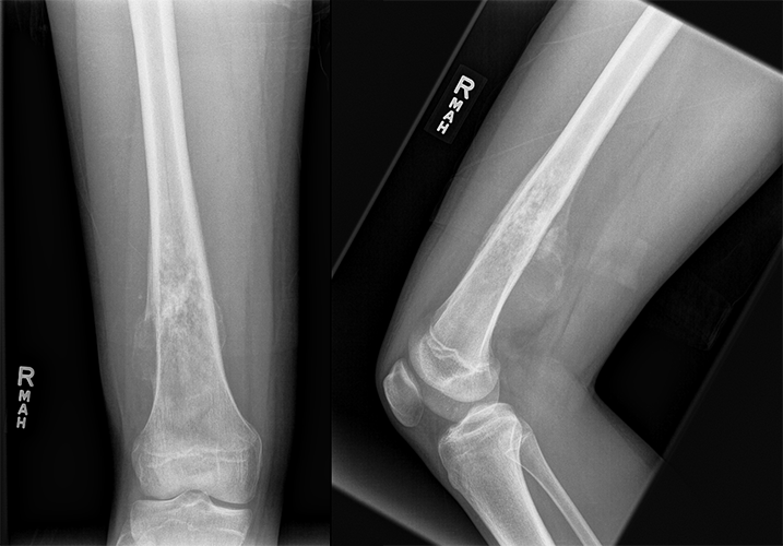
X-ray Femur
There is evidence of a malignant lesion.
The distal half of the femur contains a heterogeneous lesion resulting in medullary and cortical bone destruction with a moth-eaten appearance.
The periosteum of the femur is normal.
The characteristic finding of a Codman triangle, often seen in osteosarcoma, represents periosteal reaction of the distal aspect of the femur. A Codman triangle is a form of periosteal reaction seen when an aggressive bone lesion grows faster than the new periosteum can be formed.
View the full study if you'd like to take a look yourself.
Second Imaging Study
What is the next imaging study you will order?
CT scan
MRI is an essential tool for further characterizing bone lesions detected on radiographs, aiding in accurately grading bone tumors. It is also the preferred modality for staging bone tumors due to its superior ability to detail the relationship between the tumor and surrounding tissues. Contrast-enhanced MRI can additionally assist in distinguishing benign from malignant tumors and facilitate biopsy planning. The images are not shown for the purposes of this case.
Well done. You were correct
What is your Diagnosis now that you have seen the imaging results?
The patient’s presentation and imaging findings are consistent with an osteosarcoma.
Current Acuity
Initially, you selected and we suggested acuity.
Has your concern for this patient changed?
The patient requires routine, but expedited workup.
Assessment and Plan
Please provide your assessment and plan for this patient
In this case, we have a 16-year-old male who presents with a persistently worsening nocturnal pain in his right knee, accompanied by systemic manifestations, including unexplained fevers and weight loss. Lab investigations show elevated ESR, CRP, ALP, and LDH, indicative of an inflammatory or neoplastic process. A radiograph of his right knee reveals classic signs of osteosarcoma, notably a Codman triangle and periosteal reaction at the distal femur. Given these findings, we recommend proceeding with an MRI to further characterize the lesion and assess the extent of local invasion. Additionally, systemic staging, especially that evaluating the chest, is warranted to evaluate for potential metastasis. A definitive diagnosis through biopsy should be arranged. The patient should be referred to a multidisciplinary oncology team for further evaluation and management.
Lessons Learned:
- Osteosarcomas originate from mesenchymal stem cells, including osteoblasts within the periosteum.
- They exhibit a male predominance and two peak incidences: in the adolescent age group of 13-20 years and in adults over 65 years.
- The etiology can be categorized as primary or secondary.
- Primary osteosarcomas are most frequently observed in younger patients.
- Secondary osteosarcomas typically arise from pre-existing conditions such as Paget's disease, radiation-induced bone changes, or bone infarction.
- Certain risk factors increase the likelihood of osteosarcoma development, including prior exposure to radiation and genetic predispositions like Li-Fraumeni syndrome.
- Clinical presentation includes nocturnal pain, swelling, erythema, and reduced range of motion.
- Pathological fractures may also occur.
- Radiographically, osteosarcomas produce a characteristic "Codman triangle" or a "sunburst appearance" indicative of lytic bone lesions.
- Histopathologically, biopsy specimens reveal pleomorphic osteoblasts actively producing osteoid and woven bone matrix.
- The management of osteosarcoma involves a multidisciplinary approach.
- Severe cases may require amputation when the tumor burden threatens patient survival.
That's the end of the module! Once you've reviewed the video(s), you can click here for another case challenge.
Next

