Retake
P15) Ankle tenderness and inability to bear weight after a bicycle accident
Review the Learning Outcomes, Hx, PE and Labs, and begin the module with your Provisional Diagnosis. Keep hitting "Next" to move through the module.
Learning Outcomes
- Articulate your relationship with the consulting diagnostic radiologists in the evaluation of a pediatric patient with pain after a fall.
- Review the DDx considerations in a pediatric patient with pain after a fall.
- Identify the spectrum of imaging findings in appropriate modalities for evaluating a pediatric patient with pain after a fall.
History
A 13-year-old girl presents to the emergency department with pain and swelling in her left ankle following a fall. The pain started when she swerved on her bicycle to avoid a squirrel and lost her balance, landing on her left ankle, which inverted upon impact. She is unable to bear weight on her left foot.
Physical Exam
Vitals: BP 110/64 mmHg, HR 101 bpm, RR 18 breaths/min, Temp 98.6°F, SpO2 98.6%
Musculoskeletal: Notable swelling in the left ankle. Tenderness upon palpation over the left posterior medial malleolus. Dorsalis pedis pulses are 2+ and sensation in the foot is intact.
Labs
None.
Provisional Diagnosis
Select the Dx you believe is most appropriate
An ankle fracture is most likely in a child of this age that is unable to bear weight after a fall with significant tenderness to palpation.
Well done. You were correct
Potential Acuity
What is your assessment of the likely acuity for this patient?
Well done. You were correct
This patient requires routine, but expedited workup as their suspected condition is not immediately life or limb-threatening.
First Imaging Study
What is the first imaging study you will order?
Considering that there is tenderness at the posterior edge of the medial malleolus and inability to bear weight, the patient requires an ankle X-ray per the Ottawa ankle rules.
Well done. You were correct
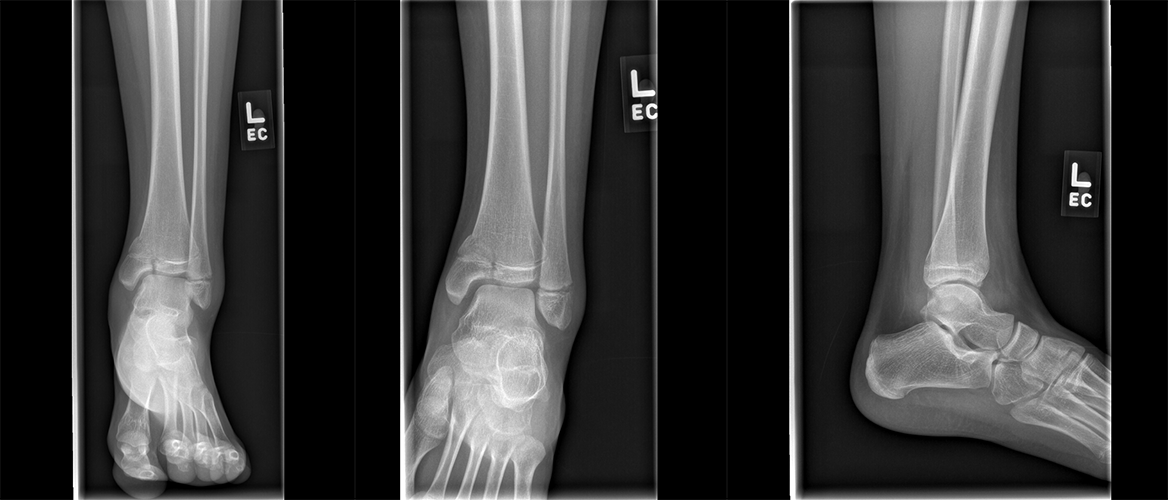
Pertinent Imaging Observations
Click on the links below to view images from the study, and assess these key findings as best you can.
AP and Lateral radiograph
There is a fracture involving the distal tibial:
The fracture extends inferolaterally into through the metaphysis, physis, and epiphysis.
The fracture can be classified as a:
Salter Harris fractures are specific to pediatric patients and involve the growth plate of the bone. This represents a Salter Harris Type IV fracture as it extends through the metaphysis, physis, and epiphysis. Type I involves the physis, Type II involves the metaphysis and physis, Type III involves the epiphysis and physis, and Type V involves compression or crushing of the growth plate.
View the full study if you'd like to take a look yourself.
Second Imaging Study
What is the next imaging study you will order?
No further imaging is required as the diagnosis is made with the X-ray.
Well done. You were correct
What is your Diagnosis now that you have seen the imaging results?
The patient has a Salter-Harris IV fracture involving the lateral distal tibia. The imaging findings are also concerning for a Salter-Harris I fracture of the left lateral malleolus of the fibula.
Current Acuity
Initially, you selected and we suggested acuity.
Has your concern for this patient changed?
The patient requires routine, but expedited workup.
Assessment and Plan
Please provide your assessment and plan for this patient
A 13-year-old girl presents with significant swelling and bruising of her left ankle. Plain radiographs demonstrated a Salter-Harris IV fracture of the left medial malleolus and a potential nondisplaced Salter-Harris I fracture of the left lateral malleolus. An orthopedist should be consulted for further management.
Lessons Learned:
- The Salter-Harris classification system is used to categorize physeal plate fractures in children, with each fracture grade determined by the extent of physis involvement.
- These fractures typically occur in active children during their growth spurts.
- The classification system assists providers in evaluating the risk of premature physeal fusion.
- Diagnosis involves X-ray, CT, and MRI imaging.
- Treatment options vary from immobilization to surgical fixation, depending on the fracture type.
- Compared to Salter-Harris I or II, Salter-Harris fractures III-V are more likely to require operative intervention and warrant an orthopedic consultation.
Socioeconomic Factors: Research suggests that children with siblings and those from higher-income families face a greater risk of fractures.
That's the end of the module! Once you've reviewed the video(s), you can click here for another case challenge.
Next

