P14) Forearm pain after fall on outstretched hand
Review the Learning Outcomes, Hx, PE and Labs, and begin the module with your Provisional Diagnosis. Keep hitting "Next" to move through the module.
Learning Outcomes
- Articulate your relationship with the consulting diagnostic radiologists in the evaluation of a pediatric patient with pain after a fall.
- Review the DDx considerations in a pediatric patient with pain after a fall.
- Identify the spectrum of imaging findings in appropriate modalities for evaluating a pediatric patient with pain after a fall.
History
Physical Exam
Labs
Provisional Diagnosis
Potential Acuity
What is your assessment of the likely acuity for this patient?
First Imaging Study
What is the first imaging study you will order?
Pertinent Imaging Observations
Click on the links below to view images from the study, and assess these key findings as best you can.
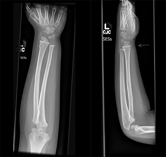
Xray Case
What besst describes the findings of the X-rays of the forearm?

Watch our video
Second Imaging Study
What is the next imaging study you will order?
What is your Diagnosis now that you have seen the imaging results?
Current Acuity
Initially, you selected and we suggested acuity.
Has your concern for this patient changed?
Assessment and Plan
Please provide your assessment and plan for this patient
Lessons Learned:
- Buckle fractures are usually stable injuries that occur due to a compressive force, and surgery is not typically necessary. They commonly occur at the distal metaphysis where the bone is most porous.
- An axial loading force from a fall on an outstretched hand is the most common cause of a buckle fracture. In contrast, greenstick fractures usually result from a rotational force.
- X-ray findings of a buckle fracture typically include buckling out of one or both cortexes of the long bone.
- Buckle fractures are generally stable and can usually be managed with a removable soft arm splint or soft elastic bandage for a few weeks to immobilize the affected area.
- Buckle fractures have an excellent prognosis, and complications are rare. Follow-up appointments or imaging are not usually necessary if the pain resolves at home.
Socioeconomic Factors: Several clinical trials have generally suggested that home care is effective, and that uncomplicated buckle fractures do not require outpatient primary care provider follow-up or imaging.
That's the end of the module! Once you've reviewed the video(s), you can click here for another case challenge.
