N8) Altered mental status, photophobia, and phonophobia
Review the Learning Outcomes, Hx, PE and Labs, and begin the module with your Provisional Diagnosis. Keep hitting "Next" to move through the module.
Learning Outcomes
- Articulate your relationship with the consulting diagnostic radiologists in the evaluation of a patient with altered mental status.
- Review the DDx considerations in a patient with altered mental status.
- Identify the spectrum of imaging findings in appropriate modalities for evaluating a patient with altered mental status.
History
Physical Exam
Labs
Provisional Diagnosis
Potential Acuity
What is your assessment of the likely acuity for this patient?
First Imaging Study
What is the first imaging study you will order?
Pertinent Imaging Observations
Click on the links below to view images from the study, and assess these key findings as best you can.
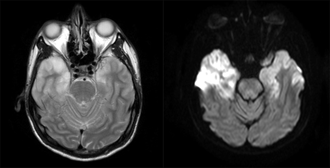
Head MRI with and without contrast
There is a space occupying lesion.
What best describes the findings on the MRI?

Watch our video
Second Imaging Study
What is the next imaging study you will order?
What is your Diagnosis now that you have seen the imaging results?
Current Acuity
Initially, you selected and we suggested acuity.
Has your concern for this patient changed?
Assessment and Plan
Please provide your assessment and plan for this patient
Lessons Learned:
- Herpes Encephalitis is the most common cause of fatal sporadic encephalitis in USA in all age groups. The most common cause is viral reactivation from HSV-1. Patients classically do not present with meningeal signs.
- CSF analysis with PCR is the gold standard for establishing diagnosis. It should be performed after a space occupying lesion is ruled out to prevent iatrogenic brain herniation. MRI will show bilateral temporal lobe hyperintensities on T2 and diffusion-weighted images.
Socioeconomic Factors: HSV-1 is common and affects up to half of the general population.
That's the end of the module! Once you've reviewed the video(s), you can click here for another case challenge.
Contributors:
Abeer Dagra, MS3 - Content Contributor
Kevin Pierre, MD - Editor
Robbie Slater, MD - Supervising Editor
Bayar Batmunh, MS - Coordinator
