Retake
Review the Learning Outcomes, Hx, PE and Labs, and begin the module with your Provisional Diagnosis. Keep hitting "Next" to move through the module.
Learning Outcomes
- Articulate your relationship with the consulting diagnostic radiologists in the evaluation of a patient with foot pain.
- Review the DDx considerations in a patient with foot pain.
- Identify the spectrum of imaging findings in appropriate modalities for evaluating a patient with foot pain.
History
A 65-year-old male complains of left heel pain over the past few months. He presents today due to inability to bear weight on the left foot. The patient reports having never seen a health care provider. He notes that he gets up to urinate several times per night.
Physical Exam
BP: 128/79 HR 105, RR 20, Temp 102.3F, O2 saturation 100%. Neuro: no sensation on bilateral plantar feet to pinprick. Deep ulcer on right heel that is 1cm in diameter. Bone is accessible with probe. The patient does not feel discomfort with the exam.
Labs
WBC: 18 x 109/L (nl: 4.5 x 10⁹/L - 11 x 109/L); Fasting blood glucose: 290 mg/dL (normal 99mg/dL); Hgb A1c: 12.2% ; UA glucose: 45mg/dL (nl 0-15mg/dL); Bone culture: pending
Provisional Diagnosis
Select the Dx you believe is most appropriate
This patient’s presentation is consistent with previously undiagnosed, uncontrolled type 2 diabetes. His physical exam is consistent with peripheral neuropathy secondary to T2DM. As such, he developed a heel pressure ulcer. Osteomyelitis of the heel should be suspected considering his fever and because the bone is able to be probed within the ulcer crater. This patient with sudden worsening of pain may also have a pathologic fracture secondary to the osteomyelitis.
Well done. You were correct
Potential Acuity
What is your assessment of the likely acuity for this patient?
Well done. You were correct
This patient meets sepsis criteria. They should undergo urgent workup.
First Imaging Study
What is the first imaging study you will order?
X-ray of the foot is the appropriate initial imaging modality for suspected foot osteomyelitis in a patient with diabetes mellitus.
Well done. You were correct
Pertinent Imaging Observations
Click on the links below to view images from the study, and assess these key findings as best you can.
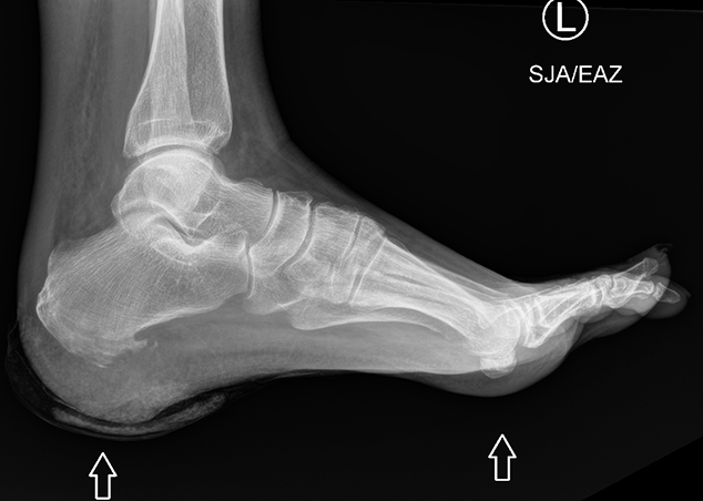
X-ray of foot
There is an identifiable fracture.
There is no sharp cortical or trabecular disruption suggesting the presence of a fracture.
There is bony erosion.
There is bony erosion suggesting osteomyelitis.
The bony erosion is at the
There is bony erosion at the plantar cortex of the calcaneus.
View the full study if you'd like to take a look yourself.
Second Imaging Study
What is the next imaging study you will order?
An MRI of the foot can confirm and determine the extent of the osteomyelitis.
Well done. You were correct
Pertinent Imaging Observations
Click on the links below to view images from the study, and assess these key findings as best you can.
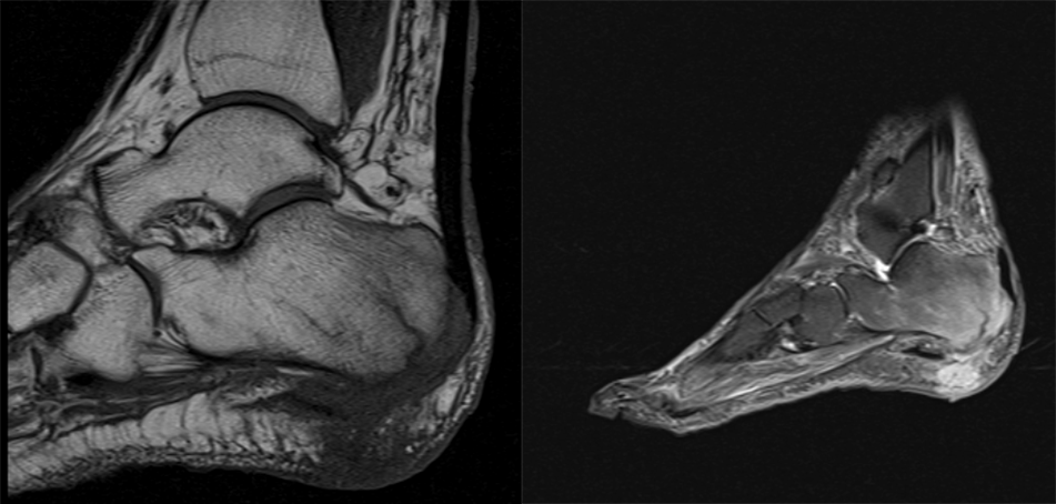
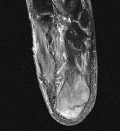
MRI of Foot
The calcaneal tuberosity is
The calcaneal tuberosity is hypointense on T1 and hyperintense on T2, suggesting inflammation and edema. These findings are consistent with osteomyelitis.
There is a fracture of the calcaneal tuberosity
There is a fracture line at the calcaneal tuberosity.
View the full study if you'd like to like a look yourself
Third Imaging Study
What is the next imaging study you will order?
No further imaging is required as the diagnosis is made with the radiograph and MRI.
What is your Diagnosis now that you have seen the imaging results?
Considering that the fracture is occurring in the setting of osteomyelitis, which compromises the bone integrity, it is considered a pathologic fracture. Furthermore, a stress fracture would appear on imaging as a sclerotic band on the radiograph or MRI.
Current Acuity
Initially, you selected and we suggested acuity.
Has your concern for this patient changed?
This patient meets sepsis criteria. They should undergo urgent workup and management.
Assessment and Plan
Please provide your assessment and plan for this patient
This is a 65-year-old male with uncontrolled diabetes mellitus presenting with a stage 4 foot ulcer and calcaneal osteomyelitis with a pathologic fracture secondary to peripheral neuropathy. The patient should be initiated on a broad-spectrum antibiotic regimen, which will be narrowed when the wound cultures return. Wound care and orthopedic surgery should be consulted for wound management. Endocrinology should be consulted for management of the patient’s diabetes. We should also ensure to set up appropriate follow-up with a primary care provider.
Lessons Learned: - Patients with uncontrolled diabetes are at risk for foot ulcers and infectious secondary to a compromised immune system and inadvertent foot damage secondary to peripheral neuropathy.
- Radiographs, which will show bony erosion, are the initial modality when foot osteomyelitis is suspected. The study can be followed by an MRI.
- All imaging findings should be reviewed as to avoid satisfaction of search. For example, this patient was also found to have plantar fasciitis with a plantar fascia tear.
Socioeconomic Factors: Lack of exercise in diabetic patients is associated with increased risk of diabetic foot problems.
That's the end of the module! Once you've reviewed the video(s), you can click here for another case challenge.
Contributors:
Kevin Pierre, MD - Editor
Robbie Slater, MD - Supervising Editor
Bayar Batmunh, MS - Coordinator
Next

