M4) Foot pain after a fall from a horse
Review the Learning Outcomes, Hx, PE and Labs, and begin the module with your Provisional Diagnosis. Keep hitting "Next" to move through the module.
Learning Outcomes
- Articulate your relationship with the consulting diagnostic radiologists in the evaluation of a patient with foot pain.
- Review the DDx considerations in a patient with foot pain.
- Identify the spectrum of imaging findings in appropriate modalities for evaluating a patient with foot pain.
History
Physical Exam
Labs
Provisional Diagnosis
Potential Acuity
What is your assessment of the likely acuity for this patient?
First Imaging Study
What is the first imaging study you will order?
Pertinent Imaging Observations
Click on the links below to view images from the study, and assess these key findings as best you can.
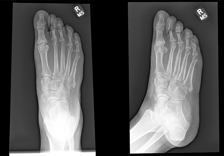
X-Ray 3-view right foot
There is appropriate alignment between each of the tarsal-metatarsal joints.
There is a metatarsal fracture.

Watch our video
Second Imaging Study
What is the next imaging study you will order?
What is your Diagnosis now that you have seen the imaging results?
Current Acuity
Initially, you selected and we suggested acuity.
Has your concern for this patient changed?
Assessment and Plan
Please provide your assessment and plan for this patient
Lessons Learned:
- The Lisfranc ligament attaches the medial cuneiform to the base of the 2nd metatarsal on the plantar aspect of the mid-foot.
- The tarsometatarsal (TMT) joint complex forms an arch that provides stability to the midfoot. The 2nd tarsometatarsal joint acts like a keystone in this arch.
- A Lisfranc injury is a complex injury involving the TMT joint complex.
- Pain to the midfoot with pronation and abduction, as well as plantar ecchymosis, should increase suspicion for Lisfranc injury.
- The first best diagnostic modality is a three views X-ray of the foot. Fractures to the tarsometatarsal joint complex increase the likelihood that the patient will require surgery.
- In cases where radiographs are normal or equivocal, but a Lisfranc injury is still suspected, an CT or MRI of the foot should be ordered.
Socioeconomic Factors:
- Bony and ligamentous injury to the foot may require surgery and significant time away from work. A meta-analysis of the socioeconomic impact of orthopedic trauma suggests that of 13% of fracture patients may lose employment because of injury.
- Patients with Lisfranc injury who underwent operative intervention may require up to a year before returning to their level of prior level of activity. Some patients are unable to return to their full level of activity.
That's the end of the module! Once you've reviewed the video(s), you can click here for another case challenge.
