C10) Pre-Operative Evaluation
Review the Learning Outcomes, Hx, PE and Labs, and begin the module with your Provisional Diagnosis. Keep hitting "Next" to move through the module.
Learning Outcomes
- Articulate your relationship with the consulting diagnostic radiologists in the evaluation of a patient with an incidental finding.
- Identify the spectrum of imaging findings in appropriate modalities for evaluating patients with incidental findings.
History
Physical Exam
Labs
Provisional Diagnosis
Potential Acuity
What is your assessment of the likely acuity for this patient?
First Imaging Study
What is the first imaging study you will order?
Pertinent Imaging Observations
Click on the links below to view images from the study, and assess these key findings as best you can.
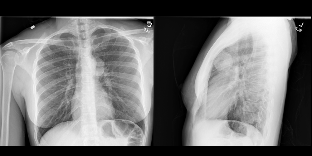
PA and Lateral Chest X-ray
What best describes the findings on the Chest X-ray?

Watch our video
Second Imaging Study
What is the next imaging study you will order?
Pertinent Imaging Observations
Click on the links below to view images from the study, and assess these key findings as best you can.
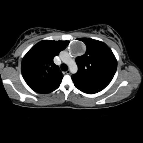
Chest CT
What best describes the findings on the Chest CT?
Watch our video
Third Imaging Study
What is the next imaging study you will order?
What is your Diagnosis now that you have seen the imaging results?
Current Acuity
Initially, you selected and we suggested acuity.
Has your concern for this patient changed?
Assessment and Plan
Please provide your assessment and plan for this patient
Lessons Learned: The most common anterior mediastinal masses in adults are teratoma, thyroid goiter, thymoma, and lymphoma.
Socioeconomic Factors:
- Indications for pre-operative chest radiography include suspected or pre-existing heart or lung conditions. Otherwise, pre-operative chest radiography should not be routinely performed in asymptomatic young or middle-aged patients undergoing surgery outside the thoracic cavity.
- It is important to carefully review all imaging regardless of indication. Early incidental detection can significantly improve outcomes.
That's the end of the module! Once you've reviewed the video(s), you can click here for another case challenge.
Contributors:
Kevin Pierre, MD - Editor
Robbie Slater, MD - Supervising Editor
Bayar Batmunh, MS - Coordinator
