Retake
C6) Hemoptysis Following Recent Travel from a Another Country
Review the Learning Outcomes, Hx, PE and Labs, and begin the module with your Provisional Diagnosis. Keep hitting "Next" to move through the module.
Learning Outcomes
- Articulate your relationship with the consulting diagnostic radiologists in the evaluation of a patient with fever and cough.
- Review the DDx considerations in fever and cough.
- Identify the spectrum of imaging findings in appropriate modalities for evaluating patients with fever and cough.
History
A 30-year-old male presents with fever, productive cough with bloody sputum, and night sweats worsening over the last month. The patient recently immigrated from Bangladesh and has recently started taking infliximab for Crohn’s disease.
Physical Exam
BP: 130/94 HR 81, RR 15, Temp 38.0, O2 saturation 99%.
General: Patient appears thin and cachectic.
Pulmonary: Crackles in right upper lung field.
HENT: Cervical lymphadenopathy.
Labs
WBC count: 12.0 × 10^9/L (normal: 4.5 to 11.0 × 10^9/L)
Provisional Diagnosis
Select the Dx you believe is most appropriate
Both a bronchogenic carcinoma and tuberculosis can present similarly with constitutional symptoms as seen here. Since the patient is taking a TNF-a inhibitor, they are at increased risk for reactivation tuberculosis had they previously contracted TB and been in the latent stage.
Well done. You were correct
Potential Acuity
What is your assessment of the likely acuity for this patient?
Well done. You were correct
Urgent workup is required. Airborne precautions should be immediately put in place if the patient has tuberculosis.
First Imaging Study
What is the first imaging study you will order?
A chest x-ray would be the best first examination. It can be completed faster than the CT scan and may strongly suggest the diagnosis.
Well done. You were correct
Pertinent Imaging Observations
Click on the links below to view images from the study, and assess these key findings as best you can.
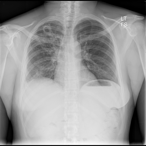
PA Chest X-ray
What best describes the findings on the PA Chest X-ray?
There is a cavitary lesion in the right lung apex with fluffy, nodular airspace opacities. A bronchogenic carcinoma can also present as a cavitating lesion. However, the opacities suggest that this is more likely an infectious process.
View the full study if you'd like to take a look yourself.
Pertinent Imaging Observations
Click on the links below to view images from the study, and assess these key findings as best you can.
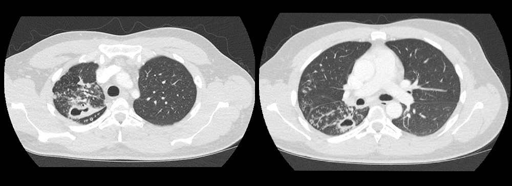
Chest CT
What best describes the findings on the Chest CT?
There is a cavitary lesion in the posterior portion of the right upper lobe. There is also endobronchial spread along nearby airways characterized by ill-defined fluffy air-space infiltrates, and sharply marginated linear branching opacities representing the tree in bud sign throughout the right lung. These findings are characteristic of tuberculosis infection.
View the full study if you'd like to like a look yourself
Third Imaging Study
What is the next imaging study you will order?
No further imaging is required.
What is your Diagnosis now that you have seen the imaging results?
The patient is presenting with post-primary tuberculosis (reactivation / secondary tuberculosis), characterized by cavitary lesions in the superior lungs, poorly defined, patchy airway opacities, and tree-in-bud sign. Primary tuberculosis would likely present with a Ghon lesion, representing a caseating granuloma (tuberculoma) that calcifies. Miliary tuberculosis would present as innumerable bilateral subcentimeter lung nodules representing disseminated, uncontrolled spread of TB.
Current Acuity
Initially, you selected and we suggested acuity.
Has your concern for this patient changed?
The patient should immediately be placed on respiratory precautions and undergo testing for tuberculosis.
Assessment and Plan
Please provide your assessment and plan for this patient
The patient is a 30-year-old recent immigrant presenting with constitutional symptoms and hemoptysis. Imaging is consistent with post-primary tuberculosis. The patient should be placed on airborne precautions undergo diagnostic tuberculosis testing. Pulmonology should be consulted and the patient should be placed on anti-tuberculous drugs once the diagnosis is confirmed.
Lessons Learned: TNF-a inhibitors increase the risk for reactivation tuberculosis. The pertinent imaging findings of tuberculosis depend on the stage: (1) Primary pulmonary tuberculosis: (a) Initial infection: Non-specific patchy consolidations that are not always detectable. (b) Infection localization: Tuberculoma (caseating granuloma) formation, then formation of a Ghon complex (tuberculoma calcification). (2) Post-primary (reactivation / secondary) tuberculosis (a) Reactivation after latent stage: Cavitary lesion(s) and signs of endobronchial spread (tree-in-bud nodules, scattered airspace opacities with indistinct borders) (3) Miliary tuberculosis (a) Widespread subcentimeter lung nodules representing disseminated, uncontrolled spread of TB when the immune system cannot clear the infection.
Socioeconomic Factors: Another reasonable clinical decision-making pathway is to immediately place the patient under airborne precautions and order diagnostic testing for tuberculosis following the X-ray. This is especially important in low-resource settings where the use of imaging is limited.
That's the end of the module! Once you've reviewed the video(s), you can click here for another case challenge.
Contributors:
Kevin Pierre, MD - Editor
Robbie Slater, MD - Supervising Editor
Bayar Batmunh, MS - Coordinator
Next

