Retake
C7) Dry Cough and Dyspnea on Exertion in a Young Female
Review the Learning Outcomes, Hx, PE and Labs, and begin the module with your Provisional Diagnosis. Keep hitting "Next" to move through the module.
Learning Outcomes
- Articulate your relationship with the consulting diagnostic radiologists in the evaluation of a patient with cough and weight loss.
- Review the DDx considerations in cough and weight loss.
- Identify the spectrum of imaging findings in appropriate modalities for evaluating patients with cough and weight loss.
History
A 29-year-old female presents with a worsening dry cough, malaise, and fever. She also now becomes short of breath when walking long distances. She endorses dry eyes and dry mouth. She also reports weight loss of 15 pounds over the past four months. She denies any smoking.
Physical Exam
BP: 135/90, HR 72, RR 21, Temp 38.0C, O2 saturation 94%. Skin: Red nodules on anterior legs that are tender to palpation. Purple rash on cheeks and nose.
Heart: S4 present.
Vision: Grossly reduced visual acuity using Snellen eye chart.
Labs
Calcium: 12mg/dL (normal 8.5-10.2mg/dL)
Provisional Diagnosis
Select the Dx you believe is most appropriate
The constitutional symptoms, dry cough, and dyspnea on exertion are concerning for sarcoidosis with pulmonary involvement. This case is also concerning for extra-pulmonary involvement, including anterior uveitis, lupus pernio, erythema nodosum, restrictive cardiomyopathy, and hypercalcemia from hypervitaminosis from 1-alpha-hydroxylase production.
Well done. You were correct
Potential Acuity
What is your assessment of the likely acuity for this patient?
Well done. You were correct
While she requires further workup, her condition is not immediately life-threatening.
First Imaging Study
What is the first imaging study you will order?
X-ray is readily available and has low radiation burden. Furthermore, it may reveal characteristic features of sarcoidosis with pulmonary involvement.
Well done. You were correct
Pertinent Imaging Observations
Click on the links below to view images from the study, and assess these key findings as best you can.
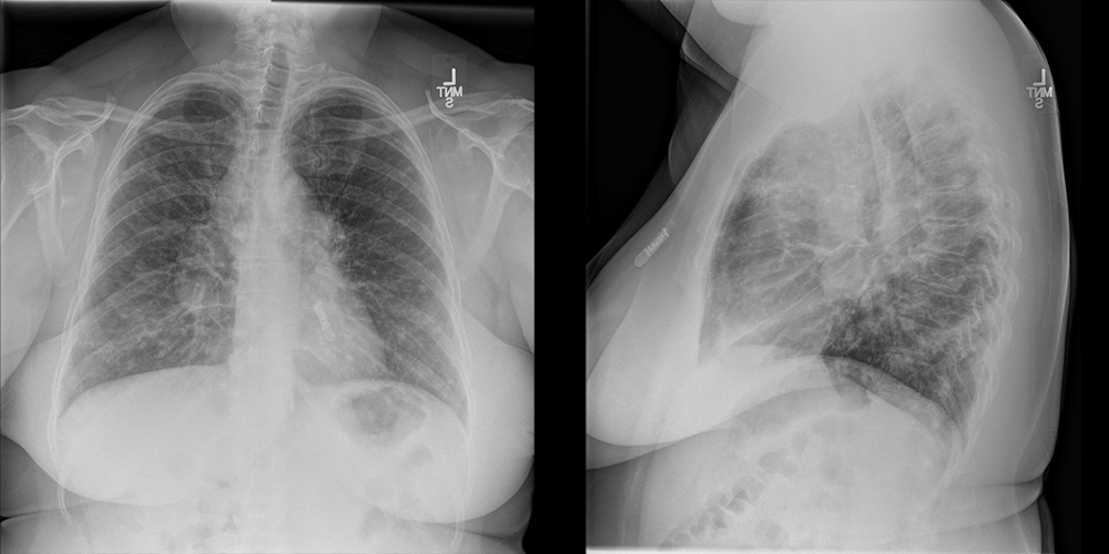
PA and Lateral Chest X-ray
What best describes the findings on the Chest X-ray?
There is bilateral hilar and right paratracheal lymphadenopathy. This reflects the “Garland triad” in sarcoidosis. These findings may represent early stage sarcoidosis or lymphoma. However, this case more likely represents sarcoidosis considering the interstitial infiltrates.
View the full study if you'd like to take a look yourself.
Second Imaging Study
What is the next imaging study you will order?
Though less sensitive than CT, the chest radiograph in this case showed characteristic manifestations of sarcoidosis for which further clinical workup is warranted.
Well done. You were correct
What is your Diagnosis now that you have seen the imaging results?
The most likely diagnosis is sarcoidosis. However, further workup is required to rule out other similar presenting diagnoses.
Current Acuity
Initially, you selected and we suggested acuity.
Has your concern for this patient changed?
Though her condition is not immediately life-threatening, the patient will require further workup for her condition.
Assessment and Plan
Please provide your assessment and plan for this patient
29-year-old female presenting with clinical symptoms and imaging concerning for sarcoidosis. While lymphoma is part of the differential, it is less likely considering the pulmonary infiltrates and other clinical findings consistent with sarcoidosis. We will obtain ACE levels and consult pulmonology for further workup with possible endoscopic lymph node biopsy.
Lessons Learned: Sarcoidosis is a disease of unclear etiology with noncaseating granulomas on biopsied tissue. It can be diagnosed and staged using chest radiographs. Stage 1) Lymphadenopathy. Stage 2) Lymphadenopathy + interstitial lung disease. Stage 3) Interstitial lung disease with shrinking / unappreciable lymph nodes. Stage 4) Advanced fibrosis. Sarcoidosis must be differentiated from lymphoma, which may present similarly. Once confirmed, sarcoidosis is treated with corticosteroids.
Socioeconomic Factors: CT is not necessary to confirm the presence of sarcoidosis when it is strongly suggested on X-ray.
That's the end of the module! Once you've reviewed the video(s), you can click here for another case challenge.
Contributors:
Kevin Pierre, MD - Editor
Robbie Slater, MD - Supervising Editor
Bayar Batmunh, MS - Coordinator
Next

