Retake
C14) Acute hypoxia and wheezing in a young female
Review the Learning Outcomes, Hx, PE and Labs, and begin the module with your Provisional Diagnosis. Keep hitting "Next" to move through the module.
Learning Outcomes
- Articulate your relationship with the consulting diagnostic radiologists in the evaluation of a patient with hypoxia.
- Review the DDx considerations in a patient with hypoxia.
- Identify the spectrum of imaging findings in appropriate modalities for evaluating a patient with hypoxia.
History
An 18-year-old female is brought in to the ED by her friends. They were jogging in cold weather when she became progressively short of breath and collapsed. The patient is unable to provide history, but her friends note she has had chronic cough, seasonal allergies, wheezing. She is given a non-rebreather, albuterol, and significantly improves. However, she still remains short of breath.
Physical Exam
BP: 132/82 HR 115, RR 30, Temp 36.1, O2 saturation 90%. General: Appears cyanotic and fatigued. Pulmonary: increased work of breathing with accessory muscle use, prolonged expiration, audible wheezing. Skin: Erythematous and scaly antecubital fossae.
Labs
ABG: pH 7.50 (nl 7.35-7.45), PaCO2 30 mmHg (35-45), PaO2 80 mmHg (nl >90).
Provisional Diagnosis
Select the Dx you believe is most appropriate
An acute asthma exacerbation is likely considering her acute dyspnea and respiratory alkalosis triggered by cold weather and exercise and history and exam suggesting atopy.
Well done. You were correct
Potential Acuity
What is your assessment of the likely acuity for this patient?
Well done. You were correct
While acute asthma exacerbation is the most likely diagnosis, further workup may rule out other pathologies as the patient did not fully respond to initial therapy.
First Imaging Study
What is the first imaging study you will order?
A chest x-ray is quickly obtainable and can rule out asthma complications like pneumothorax or pneumomediastinum.
Well done. You were correct
Pertinent Imaging Observations
Click on the links below to view images from the study, and assess these key findings as best you can.
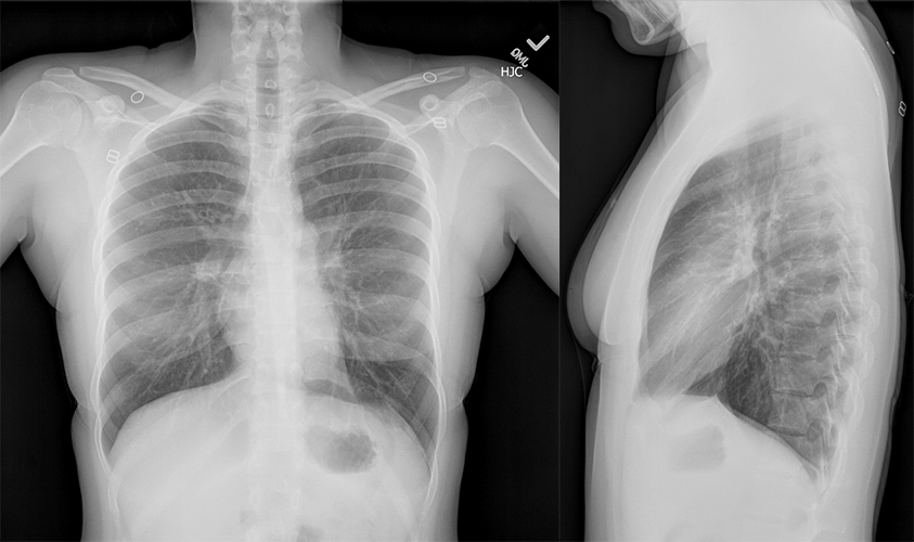
2 view chest x-ray
What best describes the findings on the Chest X-ray?
The lungs are hyperinflated – there are 10 visible posterior ribs. The heart also appears small secondary to the hyperinflation. There is also peribronchial cuffing, represented by the “donut sign” when viewing bronchi on end, and “tram tracks” when viewed tangentially.
View the full study if you'd like to take a look yourself.
Second Imaging Study
What is the next imaging study you will order?
Chest X-ray is sufficient in this case. Formal diagnosis of asthma is made with PFTs. Chest CT can assess for allergic bronchopulmonary aspergillosis (ABPA), which is not suspected in this case.
Well done. You were correct
What is your Diagnosis now that you have seen the imaging results?
The patient is in respiratory distress. She exhibited altered mental status, tachycardia, hypoxia, and cyanosis, which are signs of respiratory distress. The patient requires continued respiratory support with a low-threshold to intubate.
Current Acuity
Initially, you selected and we suggested acuity.
Has your concern for this patient changed?
The patient requires prompt treatment and close monitoring for her acute asthma exacerbation.
Assessment and Plan
Please provide your assessment and plan for this patient
The patient is a 19-year-old female presenting with respiratory distress secondary to acute asthma exacerbation with signs of impending respiratory failure. Chest X-ray supported the diagnosis of asthma and did not show a pneumothorax. She should be admitted to the ICU with a low threshold to intubate. She should later undergo PFTs to confirm and assess the severity of her asthma.
Lessons Learned:
- While findings may be unremarkable, chest X-ray may support the diagnosis of asthma. Characteristic findings include hyperinflation (10+ posterior ribs in mid-clavicular line, small cardiac silhouette, diaphragm flattening, or decreased lung markings) and peribronchial cuffing (“donut sign” when viewing bronchi on end and “tram tracks” when viewing bronchi tangentially).
Socioeconomic Factors: Asthma is diagnosed with spirometry and not imaging. However, chest x-ray should be considered when asthma complications like pneumothorax and pneumomediastinum are suspected. Chest CT can reveal ABPA when it is clinically suspected.
That's the end of the module! Once you've reviewed the video(s), you can click here for another case challenge.
Next

