Retake
C13) Dyspnea on exertion and productive cough in a long-time smoker
Review the Learning Outcomes, Hx, PE and Labs, and begin the module with your Provisional Diagnosis. Keep hitting "Next" to move through the module.
Learning Outcomes
- Articulate your relationship with the consulting diagnostic radiologists in the evaluation of a patient with cough and shortness of breath
- Review the DDx considerations in a patient with cough and shortness of breath
- Identify the spectrum of imaging findings in appropriate modalities for evaluating a patient with cough and shortness of breath
History
A 45-year-old male with 60-pack year smoking history presents to the doctor to establish medical care for the first time. Review of systems reveals worsening cough with sputum production and dyspnea on exertion over the past four years.
Physical Exam
BP: 125/78, HR 78, RR 12, Temp 37.0C, O2 saturation 89%. Pulmonary: Hyperresonance to percussion, diminished lung sounds barrel-shaped chest, expiratory wheezing, prolonged expiration. CV: Distant heart sounds. Skin: Blue fingernails and lips.
Labs
LFTs within normal limits.
Provisional Diagnosis
Select the Dx you believe is most appropriate
COPD is the most likely diagnosis considering the clinical clues for bronchitis (cough with sputum production, low oxygen, wheezing) and emphysema (hyperinflated lungs on physical exam, prolonged expiration)
Well done. You were correct
Potential Acuity
What is your assessment of the likely acuity for this patient?
Well done. You were correct
While this patient is not at immediate risk for deterioration, they require further workup.
First Imaging Study
What is the first imaging study you will order?
A chest X-ray is a good initial test in this patient with respiratory symptoms.
Well done. You were correct
Pertinent Imaging Observations
Click on the links below to view images from the study, and assess these key findings as best you can.
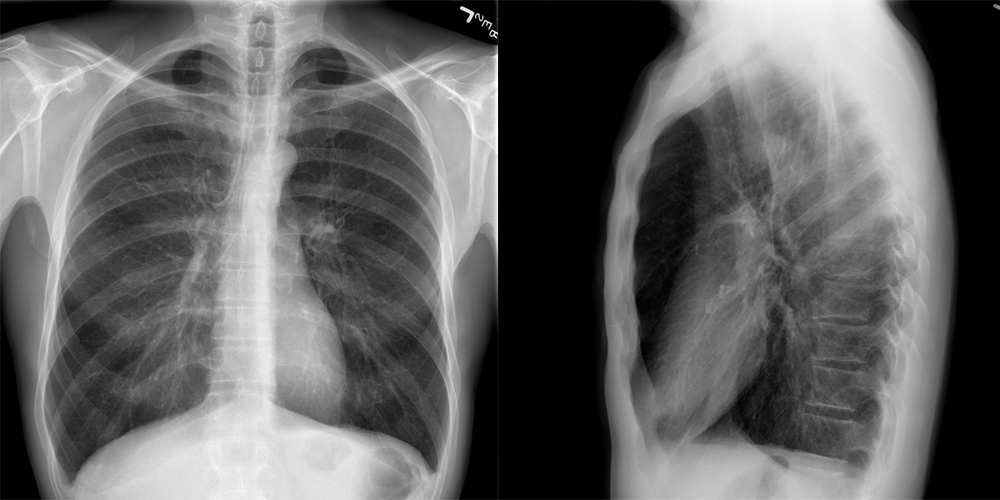
Chest X-ray
What best describes the findings on the Chest X-ray?
The patient has hyperinflated lungs (10+ posterior rib shadows, flattened diaphragms, narrow cardiomediastinal silhouette) and hyperlucent lungs consistent with COPD.
View the full study if you'd like to take a look yourself.
Second Imaging Study
What is the next imaging study you will order?
The diagnosis of COPD is strongly suggested with the CXR and can be confirmed with spirometry. However, the patient will require a low-dose screening chest CT in 5 years when he turns 50 (USPSTF guideline), which will likely show bronchial wall thickening and upper lobe centrilobular emphysema.
Well done. You were correct
What is your Diagnosis now that you have seen the imaging results?
The patient’s extensive smoking history is a risk factor for the development of their COPD. Alpha-1 antitrypsin deficiency is unlikely considering the normal LFTs and age of onset.
Current Acuity
Initially, you selected and we suggested acuity.
Has your concern for this patient changed?
The patient requires routine management.
Assessment and Plan
Please provide your assessment and plan for this patient
50-year-old male with significant smoking history presenting with clinical and imaging findings concerning for COPD. The patient will require spirometry for a definitive diagnosis. He will require counseling on smoking cessation. We will ensure he is up to date on his influenza and pneumococcal vaccines and give him a trial of a short acting beta agonist.
Lessons Learned: - COPD findings on chest x-ray include hyperinflated lungs (10+ posterior rib shadows, narrow cardiomediastinal silhouette, enlarged AP diameter, and a flattened diaphragm) and lung hyperlucency.
- Definitive diagnosis of COPD is made with spirometry revealing an FEV1/FVC ratio <0.7.
Socioeconomic Factors: Chest X-ray is not necessary to diagnose COPD. The diagnosis can be made with physical examination and incentive spirometry alone.
That's the end of the module! Once you've reviewed the video(s), you can click here for another case challenge.
Next

