Retake
C18) Severe dyspnea and hypoxia after MVC
Review the Learning Outcomes, Hx, PE and Labs, and begin the module with your Provisional Diagnosis. Keep hitting "Next" to move through the module.
Learning Outcomes
- Articulate your relationship with the consulting diagnostic radiologists in the evaluation of a patient with hypoxia.
- Review the DDx considerations in a patient with hypoxia.
- Identify the spectrum of imaging findings in appropriate modalities for evaluating a patient with hypoxia.
History
A 78-year-old male presents as a trauma alert following a motor vehicle crash. Due to prolonged extrication, he presents 2 hours following the initial injury. En route, he complained of severe right-sided chest pain and back pain. On arrival, he was intubated and mechanically ventilated due to severe dyspnea, hypopnea, and hypoxia.
Physical Exam
BP: 108/68, HR 104, RR 22, O2 saturation 87%. Pulmonary: Decreased breath sounds and dullness to percussion throughout the right lung. Crepitus to right lateral chest wall. Neuro: Full strength and sensation to light touch in all extremities.
Labs
Hgb: 11 gm/dL (nl 14-17.5gm/dL)
Provisional Diagnosis
Select the Dx you believe is most appropriate
Hemothorax secondary to intercostal injury from rib fractures is the most likely diagnosis considering the chest wall crepitus, decreased breath sounds, hypoxia, and anemia in the setting of trauma.
Well done. You were correct
Potential Acuity
What is your assessment of the likely acuity for this patient?
Well done. You were correct
This patient requires urgent workup for the cause of their respiratory failure.
First Imaging Study
What is the first imaging study you will order?
Chest X-ray is readily available in the trauma bay and should be used first.
Well done. You were correct
Pertinent Imaging Observations
Click on the links below to view images from the study, and assess these key findings as best you can.
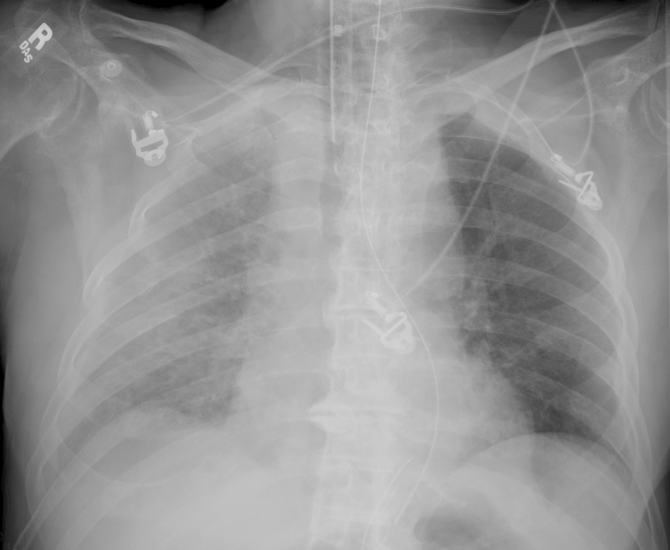
AP chest x-ray
What best describes the findings on the Chest X-ray?
There is a single right lower lateral rib fracture. The right hemithorax is opacified compared to the left, possibly representing a hemothorax or lung contusion. The mediastinum is widened. The NGT is looped in the distal esophagus.
View the full study if you'd like to take a look yourself.
Second Imaging Study
What is the next imaging study you will order?
Chest X-rays do not reliably detect rib fractures, particularly if they are nondisplaced. They can be best evaluated on CT. Furthermore, CT is the next step per ATLS protocol. It may detect more rib fractures, flail chest (3+ contiguous ribs fractures in 2+ places), the severity of rib fractures, and the presence of an actively bleeding intercostal artery contributing to the hemothorax. It will also rule out acute aortic injury as the cause of the widened mediastinum.
Well done. You were correct
Pertinent Imaging Observations
Click on the links below to view images from the study, and assess these key findings as best you can.
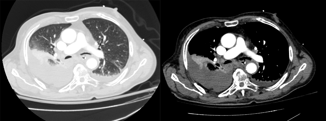
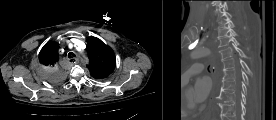
Chest CT
What best describes the findings on the Chest CT?
There is a moderate right sided hemothorax with fractures in several right lateral ribs. There is no active contrast extravasation from the intercostal artery in this case.
There is evidence of aortic injury
There is no aortic injury. The widened mediastinum on x-ray is secondary to the mediastinal hematoma from a T3-T4 distraction injury. There is also a T8 vertebral body fracture.
View the full study if you'd like to like a look yourself
Third Imaging Study
What is the next imaging study you will order?
MRI is the next step to evaluate the thoracic spine. However, the patient first requires management of their hemothorax. In the interim, they should be placed in spinal precautions (laid flat, log roll only).
What is your Diagnosis now that you have seen the imaging results?
While the patient has several contiguous rib fractures, each rib is only fractured at one location. Fractures of three contiguous ribs in two locations in each rib would predispose the patient to having flail chest.
Current Acuity
Initially, you selected and we suggested acuity.
Has your concern for this patient changed?
The patient requires chest tube drainage of their hemothorax.
Assessment and Plan
Please provide your assessment and plan for this patient
The patient is presenting with hemothorax and rib fractures. We will prepare for right chest tube insertion. We will also obtain an MRI and consult neurosurgery for the T-spine injuries.
Lessons Learned:
- A pleural effusion in the setting of trauma with associated rib fractures almost certainly represents a hemothorax.
- A hemothorax may present as an opacified hemithorax on chest x-ray in the supine trauma patient secondary to the blood layering in the posterior thoracic cavity.
- It is important to look for active extravasation from an intercostal artery, as this may require intervention.
- Always ensure appropriate placement of lines and tubes.
Socioeconomic Factors: Ultrasound as part of the eFAST may diagnose a hemothorax.
That's the end of the module! Once you've reviewed the video(s), you can click here for another case challenge.
Next

