Retake
A29) Painless hematochezia in a 75-year-old male
Review the Learning Outcomes, Hx, PE and Labs, and begin the module with your Provisional Diagnosis. Keep hitting "Next" to move through the module.
Learning Outcomes
- Articulate your relationship with the consulting diagnostic radiologists in the evaluation of a patient with hematochezia.
- Review the DDx considerations in a patient with hematochezia.
- Identify the spectrum of imaging findings in appropriate modalities for evaluating a patient with hematochezia.
History
A 75-year old male presents to the emergency department due to painless bright red blood per rectum for the past 2 days. He endorses a history of diverticulosis diagnosed during a routine colonoscopy and chronic constipation. He denies changes in stool caliber, weight loss, or abdominal pain.
Physical Exam
Vital Signs: BP 110/70, HR 86, RR 18, Temp 98.6°F, O2 saturation 97%.
Abdominal: Normal bowel sounds. No abdominal distension, pain, rebound tenderness, guarding, and abdominal bruits.
Rectal: Normal rectal tone. No palpable masses or irregularities along the mucosal wall. No external hemorrhoids or anal fissures observed. Presence of blood in rectum confirmed. Positive stool guaiac test.
Labs
RBC: 4.5 x 10^6/mm3 (N: 4.3 - 5.9 x 10^6/mm3),
Hb (g/dL): 13.2 g/dL (N: 13.5 – 17.5 g/dL),
Hct (%): 42% (N: 41% - 53%),
MCV: 80 (N: 80 - 100),
Plt: 310 x 10^3/mm3 (N: 150 – 400 x 10^3/mm3),
WBC: 5,000/mm3 (N: 4,500 – 11,000/mm3).
Provisional Diagnosis
Select the Dx you believe is most appropriate
Given the patient's age, painless hematochezia, and a history of diverticulosis and constipation, bleeding diverticulosis seems to be the most consistent diagnosis.
Well done. You were correct
Potential Acuity
What is your assessment of the likely acuity for this patient?
Well done. You were correct
The patient is hemodynamically stable and his condition is not immediately life-threatening. A routine workup, but expedited workup is recommended.
First Imaging Study
What is the first imaging study you will order?
A CT scan of the abdomen and pelvis with and without IV contrast is a sensitive and specific modality for locating the origin and understanding the nature of lower GI bleeding, even for lesions with slow bleeding rates.
Well done. You were correct
Pertinent Imaging Observations
Click on the links below to view images from the study, and assess these key findings as best you can.
CT abdomen and pelvis with and without IV contrast
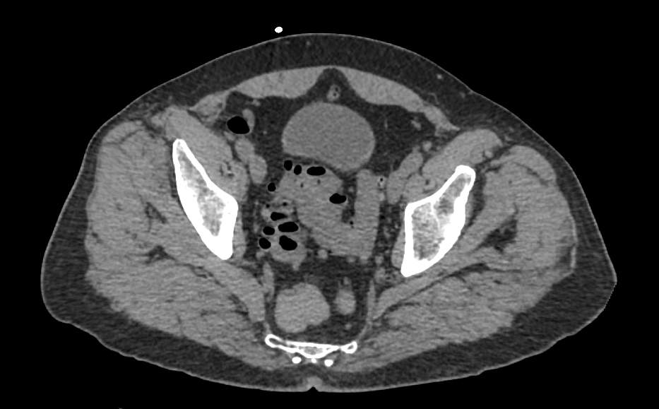
The diverticulosis is seen in the:
There is extensive diverticulosis, primarily located in the sigmoid colon.
Which phase is this image?
There is no contrast within the vasculature, indicating that this is a non-contrasted image.
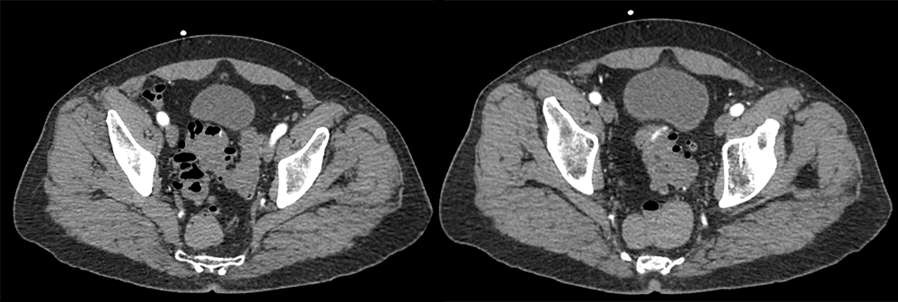
Which phase are these images?
These images appear to be captured during the arterial phase, as evidenced by the brightly enhanced iliac arteries.
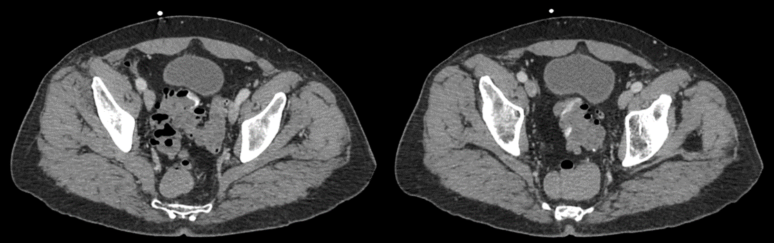
Which phase are these images?
The brightness of the arteries in these images is not as pronounced as in the previous set, suggesting these images were taken during the venous phase.
There is evidence of bleeding.
Yes, there is evidence of bleeding. The images display luminal contrast extravasation from the sigmoid diverticula, with subsequent venous pooling observed in the venous phase. This pattern is indicative of active bleeding.
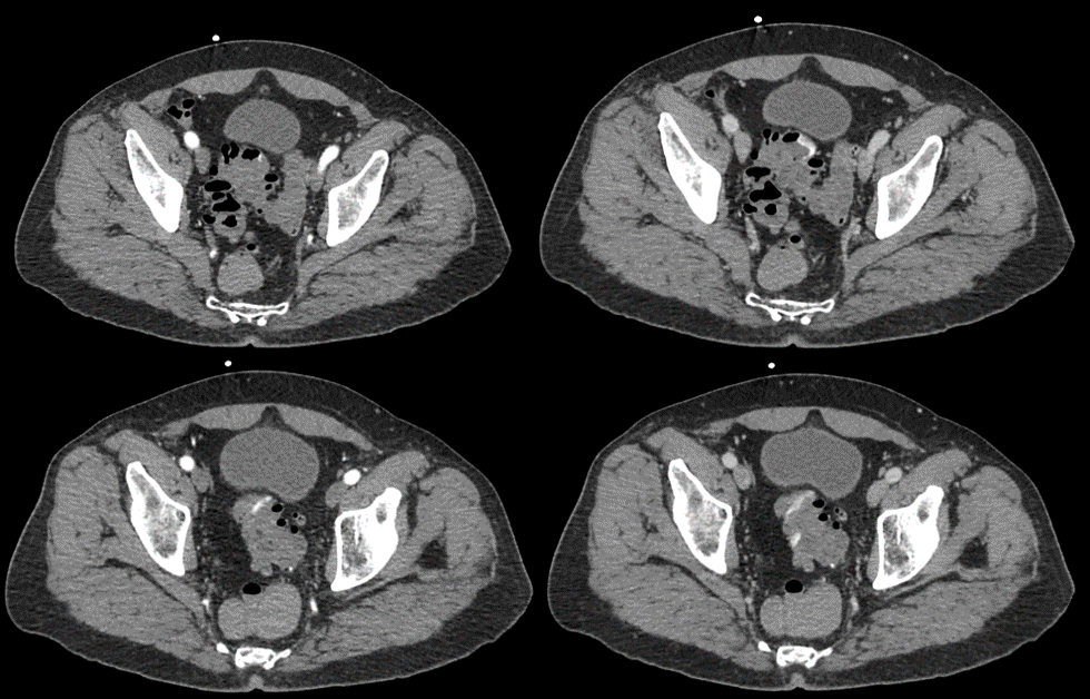
View the full study if you'd like to take a look yourself.
Second Imaging Study
What is the next imaging study you will order?
No further imaging is necessary as the diagnosis is confirmed with the diagnosis.
Well done. You were correct
What is your Diagnosis now that you have seen the imaging results?
The patient’s presentation and imaging findings are consistent with a diagnosis of bleeding diverticulosis of the sigmoid colon.
Current Acuity
Initially, you selected and we suggested acuity.
Has your concern for this patient changed?
The patient requires routine, but expedited workup.
Assessment and Plan
Please provide your assessment and plan for this patient
This 75-year-old male with a history of chronic constipation presented with painless hematochezia. A CT scan of the abdomen and pelvis confirmed bleeding sigmoid diverticulosis. A gastroenterology consultation is recommended for potential colonoscopy and endoscopic therapy. Anticoagulants or antiplatelet medications should be reviewed and possibly discontinued. The patient should be provided with stool softeners and dietary advice to prevent constipation. Hemoglobin levels should be closely monitored throughout his hospital stay.
Lessons Learned:
- In the US, diverticulosis is prevalent in about 50% of individuals over the age of 60, with chronic constipation as a major risk factor.
- Diverticulosis is the leading cause of lower GI bleeding in adults, affecting approximately 5% of individuals diagnosed with the condition.
- Diverticular bleeding results from the rupture of vasa recta, the small blood vessels near the diverticula.
- CT scanning is the initial imaging modality of choice for identifying the location and cause of bleeding. Other imaging modalities include endoscopy, nuclear medicine studies, and angiography.
- Active GI bleeding is characterized by extravasation of contrast material in the bowel lumen, which is visible during the arterial phase, with subsequent venous pooling on the delayed venous phase.
- Noncontrast images can help confirm the diagnosis of active bleeding by ruling out the presence of hyperattenuating fecal material or contrast from a previous scan.
Socioeconomic Factors: Lifestyle and environmental factors contribute to the development of diverticulosis, including a diet that is low in fiber and high in fat, obesity, low physical activity, older age, and smoking.
That's the end of the module! Once you've reviewed the video(s), you can click here for another case challenge.
Next

