Retake
A25) Jaundice and unintentional weight loss
Review the Learning Outcomes, Hx, PE and Labs, and begin the module with your Provisional Diagnosis. Keep hitting "Next" to move through the module.
Learning Outcomes
- Articulate your relationship with the consulting diagnostic radiologists in the evaluation of a patient with jaundice.
- Review the DDx considerations in a patient with jaundice.
- Identify the spectrum of imaging findings in appropriate modalities for evaluating a patient with jaundice.
History
A 75-year-old female with an 80 pack-year smoking history presents to her primary care provider with a progressive onset of jaundice, fatty stools, and unintentional weight loss of 40 pounds over the past 8 months.
Physical Exam
BP: 132/93, HR 73, RR 18, Temp 97.2, O2 saturation 99%. General: Scleral icterus. Jaundice. Abdomen: Soft, non-tender, nondistended.
Labs
CA 19-9: 500 U/mL (elevated).
Aspartate Aminotransferase (AST): 70 U/L (reference range: 10-40 U/L),
Alanine Aminotransferase (ALT): 75 U/L (reference range: 10-45 U/L),
Alkaline Phosphatase (ALP): 590 U/L (reference range: 40-130 U/L),
Total bilirubin: 7 mg/dL (reference range: 0.2-1.2 mg/dL),
Direct bilirubin: 6.5 mg/dL (reference range: 0.0-0.3 mg/dL),
Gamma-Glutamyl Transferase (GGT): 550 U/L (reference range: 9-64 U/L).
Amylase: 450 U/L (reference range: 25-125 U/L),
Lipase: 400 U/L (reference range: 10-140 U/L).
Provisional Diagnosis
Select the Dx you believe is most appropriate
The patient's risk factors, including advanced age and a significant smoking history, combined with symptoms such as jaundice, fatty stools, and unintentional weight loss, raise concerns for pancreatic adenocarcinoma. The cholestatic pattern observed in the liver function tests, along with elevated pancreatic enzymes, indicates that the potential malignancy may be causing obstructive cholestasis.
Well done. You were correct
Potential Acuity
What is your assessment of the likely acuity for this patient?
Well done. You were correct
The patient requires urgent workup and management.
First Imaging Study
What is the first imaging study you will order?
Contrast-enhanced CT is the preferred imaging technique for the diagnosis and staging of pancreatic cancer at due to its speed, robustness, and good spatial resolution, particularly for assessing tumor involvement with vascular structures. With comparable sensitivities and specificities to 3T MRI for determining resectability, CT is often chosen for its ability to optimize arterial and portal venous phase enhancement, providing a clearer visualization of the primary tumor and potential liver metastases. In this case, the patient was scanned both with and without contrast.
Well done. You were correct
Pertinent Imaging Observations
Click on the links below to view images from the study, and assess these key findings as best you can.
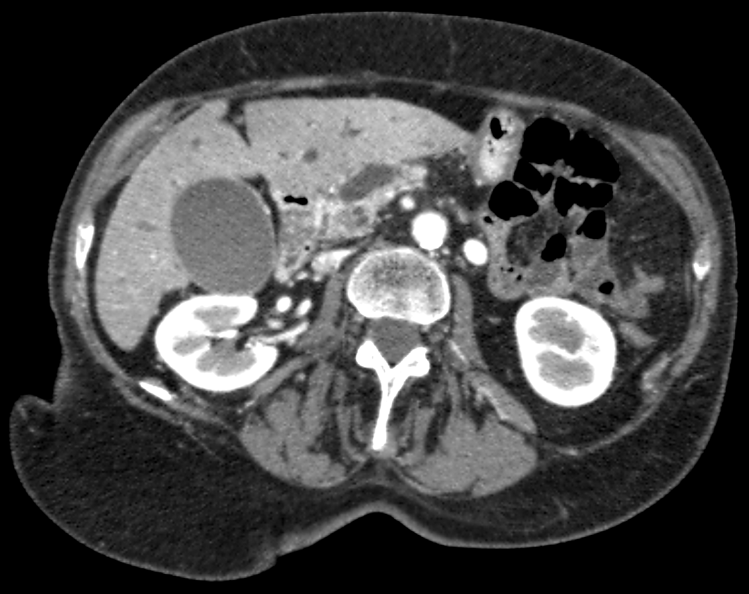
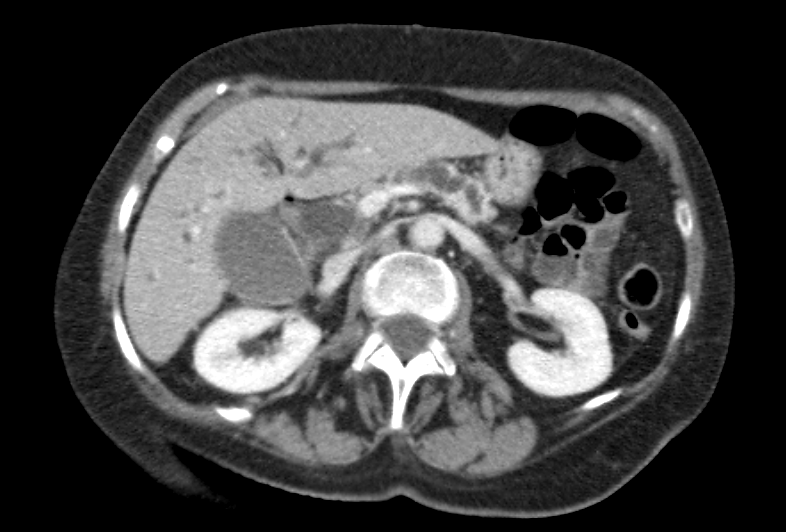
CT abdomen with and without IV contrast
There is evidence of acute pancreatitis.
There is no pancreatic edema, peripancreatic fat stranding, or fluid collection around the pancreas to suggest acute pancreatitis.
The pancreas shows:
There is a hypodense mass at the pancreatic head, which is concerning for a pancreatic malignancy, such as an adenocarcinoma.
The biliary ducts are:
The intrahepatic and extrahepatic biliary ducts are dilated.
View the full study if you'd like to take a look yourself.
Second Imaging Study
What is the next imaging study you will order?
No further imaging is required since the CT with and without IV contrast provides adequate information for the diagnosis and abdominal staging of pancreatic adenocarcinoma.
Well done. You were correct
What is your Diagnosis now that you have seen the imaging results?
The tumor may be resectable, as there is no evidence of local invasion into major blood vessels (SMA, SMV, celiac axis, or portal vein) or extensive lymph node involvement on imaging.
Current Acuity
Initially, you selected and we suggested acuity.
Has your concern for this patient changed?
The patient requires urgent workup and management.
Assessment and Plan
Please provide your assessment and plan for this patient
This is a 75-year-old female presenting with clinical and imaging findings consistent with pancreatic adenocarcinoma causing obstructive cholestasis and pancreatic ductal dilation. The tumor appears potentially resectable, given the absence of local invasion into major blood vessels (SMA, SMV, celiac axis, or portal vein) or extensive lymph node involvement. Additional imaging, such as a chest CT, should be obtained to evaluate distant metastases. The decision for surgery will depend on the presence of distant metastases and the patient's overall health and goals of care. Consultation with surgical oncology is recommended.
Lessons Learned:
- Pancreatic adenocarcinoma is the second most common gastrointestinal malignancy in the US, with a poor prognosis due to its late presentation.
- Risk factors include older age, cigarette smoking, type 2 diabetes mellitus, and genetic syndromes, among others.
- A majority of patients (around 80%) present with regional spread or metastatic disease at diagnosis, making only 15-20% eligible for potentially curative surgical treatment.
- Contrast-enhanced CT is the go-to initial imaging modality for diagnosis and staging of pancreatic cancer, as it effectively assesses tumor involvement with vascular structures, regional lymph nodes, and distant metastases, which are crucial for determining resectability.
- For borderline resectable cases, imaging helps delineate the extent of tumor abutment with visceral arteries or short-segment occlusion of certain veins, guiding the consideration of neoadjuvant therapy for downstaging.
- Additional imaging, such as chest CT, may be required during the initial workup to assess distant metastases, influencing the decision for surgery.
- Pancreaticoduodenectomy (Whipple procedure) is the standard operation for potentially resectable pancreatic cancers within the head or uncinate process.
Socioeconomic Factors: Patients with pancreatic adenocarcinoma of lower socioeconomic status are less likely to be offered surgery and, if given the option, less likely to choose surgery compared to those with higher socioeconomic status.
That's the end of the module! Once you've reviewed the video(s), you can click here for another case challenge.
Next

