A12) Hematemesis and chest pain
Review the Learning Outcomes, Hx, PE and Labs, and begin the module with your Provisional Diagnosis. Keep hitting "Next" to move through the module.
Learning Outcomes
- Articulate your relationship with the consulting diagnostic radiologists in the evaluation of a patient with chest pain.
- Review the DDx considerations in a patient with chest pain.
- Identify the spectrum of imaging findings in appropriate modalities for evaluating a patient with chest pain.
History
Physical Exam
Labs
Provisional Diagnosis
Potential Acuity
What is your assessment of the likely acuity for this patient?
First Imaging Study
What is the first imaging study you will order?
Pertinent Imaging Observations
Click on the links below to view images from the study, and assess these key findings as best you can.
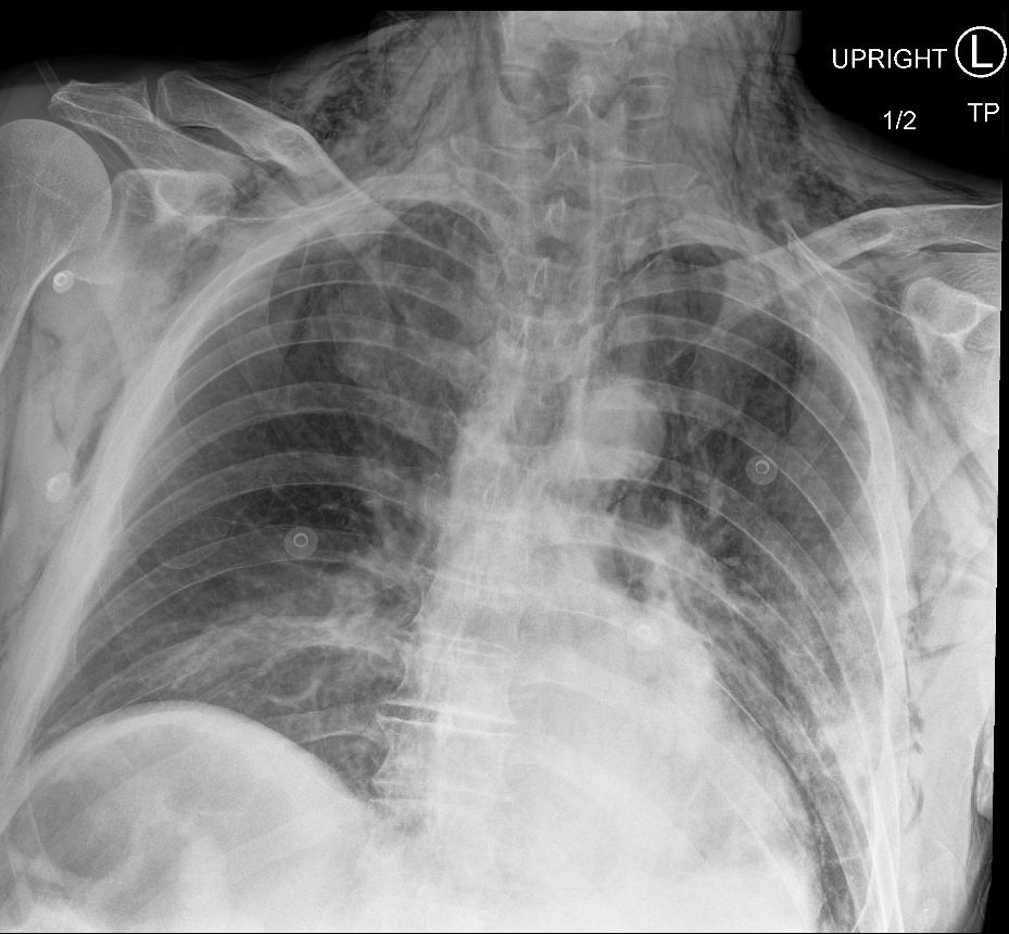
CXR
What best describes the findings on the CXR?

Watch our video
Second Imaging Study
What is the next imaging study you will order?
Pertinent Imaging Observations
Click on the links below to view images from the study, and assess these key findings as best you can.
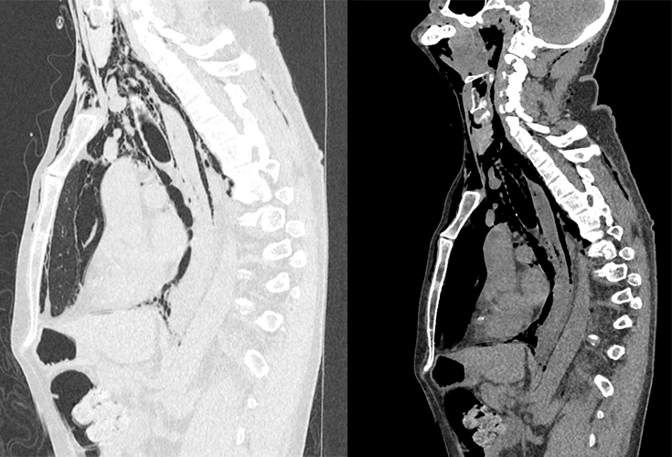
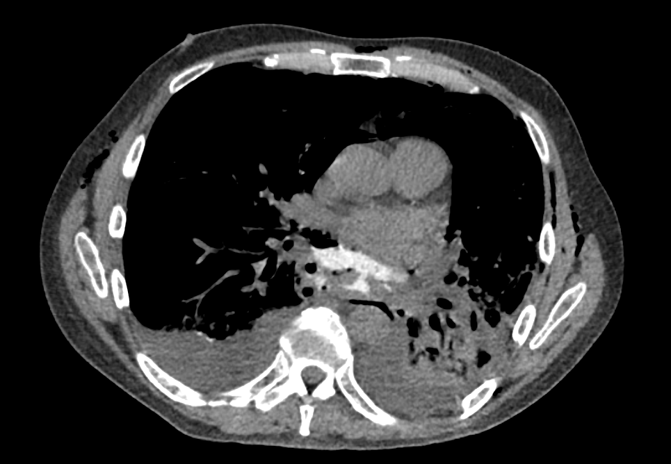
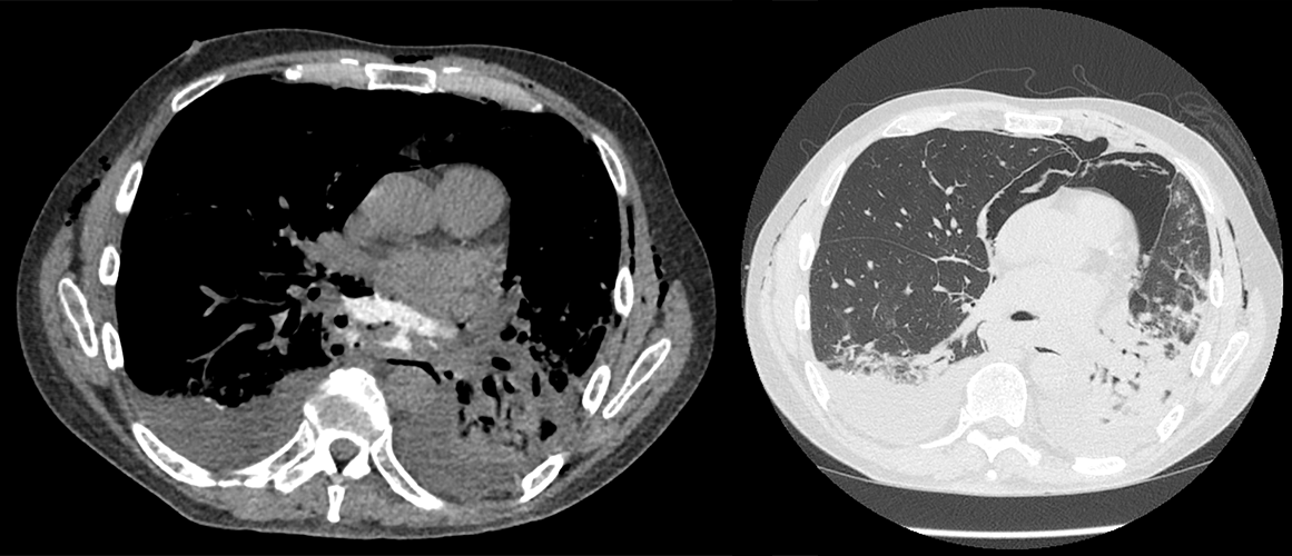
CT esophagram
There is pneumomediastinum.
The orally administered contrast:
The chest CT is otherwise normal.
Watch our video
Third Imaging Study
What is the next imaging study you will order?
What is your Diagnosis now that you have seen the imaging results?
Current Acuity
Initially, you selected and we suggested acuity.
Has your concern for this patient changed?
Assessment and Plan
Please provide your assessment and plan for this patient
Lessons Learned:
- Boerhaave syndrome can occur due to an esophageal endoscopy and balloon dilation and severe vomiting.
- Patients presenting with esophageal rupture usually have an underlying, long-standing esophageal disorder.
- Boerhaave syndrome should be suspected in a patient with severe chest, neck, or abdominal pain following severe forceful emesis. It sometimes presents as the “Mackler triad” of vomiting, chest pain, and subcutaneous emphysema. It can also lead to sepsis.
- A chest X-ray can reveal pneumomediastinum and subcutaneous emphysema.
- A CT esophagram can confirm the diagnosis by demonstrating extravasation of contrast from the esophagus.
Socioeconomic Factors: In a case such as this one where the diagnosis is strongly suspected, a CT prior to a chest X-ray is also an appropriate decision-making pathway.
That's the end of the module! Once you've reviewed the video(s), you can click here for another case challenge.
