Retake
A1) Pre-operative transplant donor evaluation
Review the Learning Outcomes, Hx, PE and Labs, and begin the module with your Provisional Diagnosis. Keep hitting "Next" to move through the module.
Learning Outcomes
- Articulate your relationship with the consulting diagnostic radiologists in the evaluation of a patient with incidental findings.
- Identify the spectrum of imaging findings in appropriate modalities for evaluating a patient with an incidental finding.
History
A 32-year-old female presents to the transplant surgery clinic for a preoperative visit for a donor nephrectomy. She would like to donate a kidney to her sister who has end-stage renal disease. She denies any past medical history. The patient has already had a pre-operative chest X-ray prior to this visit that was negative.
Physical Exam
BP: 128/71 (previous visit). Current visit: BP 125/70, HR 73, RR 17, Temp 36.4C, O2 saturation 100%. BMI 24.5.
No significant findings on physical exam.
Labs
CBC, Urinalysis, EKG, metabolic testing, HbA1C, and PT/INR within normal limits.
Donor blood type: A. Recipient blood type: A.
Crossmatch: no evidence of preformed anti-donor antibodies.
Transmissible infectious disease screening negative.
Provisional Diagnosis
Select the Dx you believe is most appropriate
Not Applicable - This module pertains to pre-transplant workup and does not involve a provisional diagnosis.
Well done. You were correct
Potential Acuity
What is your assessment of the likely acuity for this patient?
Well done. You were correct
The patient requires routine workup.
First Imaging Study
What is the first imaging study you will order?
The Organ Procurement and Transplant Network (OPTN) requires an abdominal CT, which allows for evaluation for any structural abnormalities, stones, including congenital absence of a kidney. The use of IV contrast allows for evaluation of the renal vasculature, including assessment of anatomical variants and patency, for pre-operative planning. The echocardiogram is not required. There are no findings in this case requiring further cardiac evaluation.
Well done. You were correct
Pertinent Imaging Observations
Click on the links below to view images from the study, and assess these key findings as best you can.
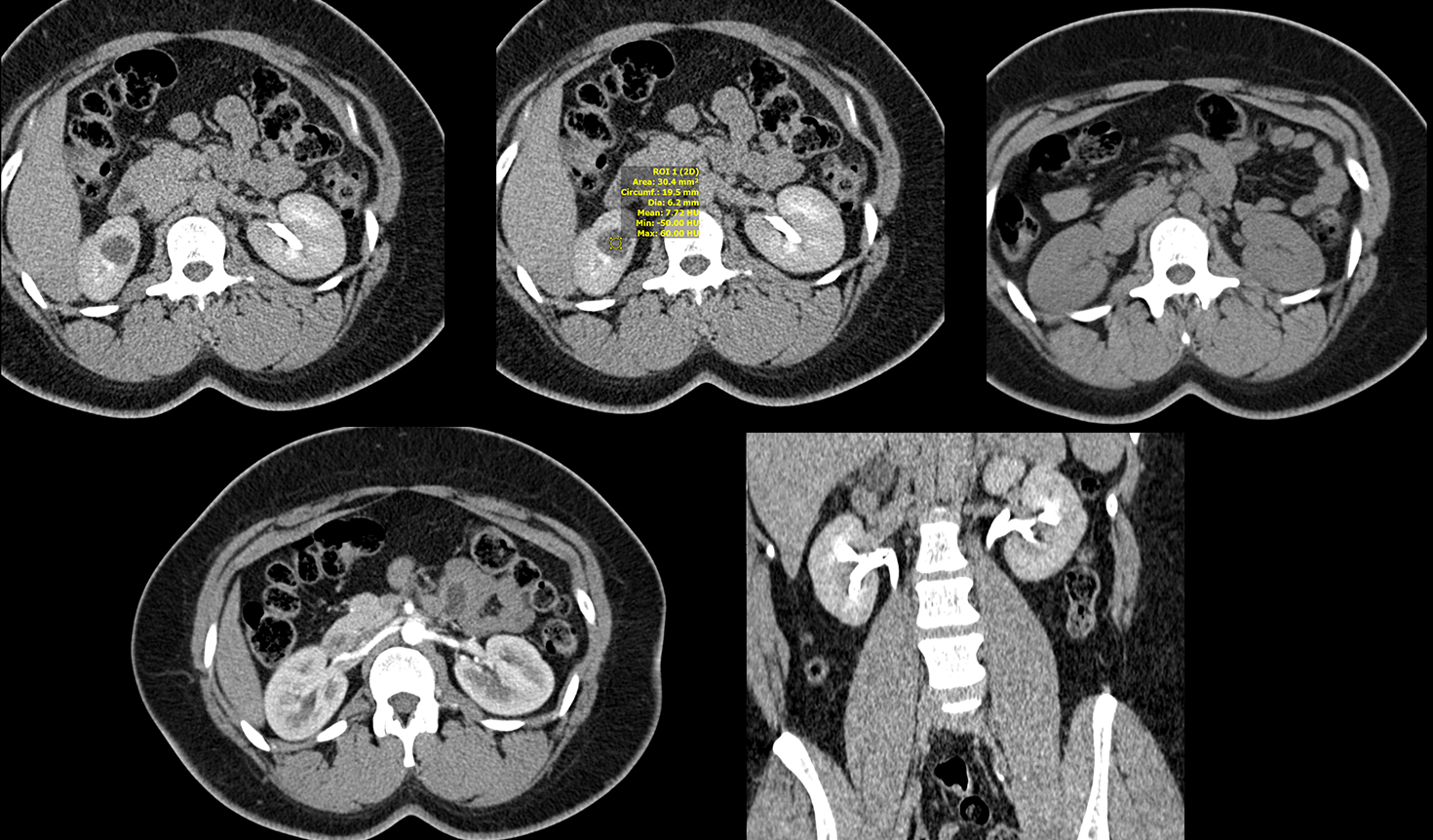
Abdominal CT with and without IV Contrast
What best describes the findings of the abdominal CT with IV contrast?
There is a right renal cystic lesion containing simple fluid without internal septations or calcifications. There is no evidence of renal stones or hydronephrosis; the renal pelvises are normal in caliber and there is no blunting of the calyces.
View the full study if you'd like to take a look yourself.
Second Imaging Study
What is the next imaging study you will order?
This simple cyst is benign and does not require further imaging workup.
Well done. You were correct
What is your Diagnosis now that you have seen the imaging results?
This represents a Bosniak I cyst as it has water density 0-20 with thin margins. A Bosniak II cyst would also have a few thin septations and punctate calcifications. Bosniak IIF cyst have multiple septa with nodular/irregular calcifications. Bosniak III cysts also have thick walls, irregular, heterogeneous septa, measurarble enhancement, and irregular calcifications. Lastly, Bosniak IV cysts have solid, soft tissue components. Malignancy risk: Bosniak I: 0%; II: 0%; IIF: ~5%; III: ~55%; IV: ~100%.
Current Acuity
Initially, you selected and we suggested acuity.
Has your concern for this patient changed?
The patient has no immediately life-threatening condition. However, the surgical team should be notified of the cyst as it may impact the patient’s candidacy for transplant.
Assessment and Plan
Please provide your assessment and plan for this patient
This patient is a 32-year-old female presenting for donor kidney evaluation. CT with and without IV contrast revealed a large Bosniak I cyst. While this cyst is benign, it is large and the surgical team should be notified of its presence as it may affect the patient’s candidacy for renal transplant.
Lessons Learned:
- Pre-operative donor kidney transplant workup includes a chest x-ray. CT of the abdomen with and without IV contrast is also required to evaluate for renal and vascular abnormalities that may affect donor transplant candidacy and surgical approach.
- Renal cysts are categorized with the Bosniak classification. Bosniak I and II cysts are benign. Bosniak IIF, III, and IV cysts require follow-up or intervention as they may be malignant.
Socioeconomic Factors: Recipients of kidney transplants from living donors generally have improved outcomes compared to recipients of kidney transplants from deceased donors.
That's the end of the module! Once you've reviewed the video(s), you can click here for another case challenge.
Next

