Ocular and Orbital Inflammation Conditions
Ocular and Orbital Inflammation Conditions
Search Pattern Assist ?History
Exam
MRI- T1 and T2 weighted images were done in the axial coronal plane with high-resolution techniques focused on the orbits and anterior and visual pathways. These include fat-suppressed and images following the intravenous administration of a paramagnetic contrast agent.
Prior Study
Findings
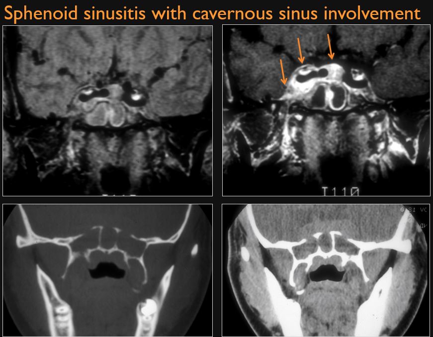
Orbit, sinonasal, cavernous and skull base
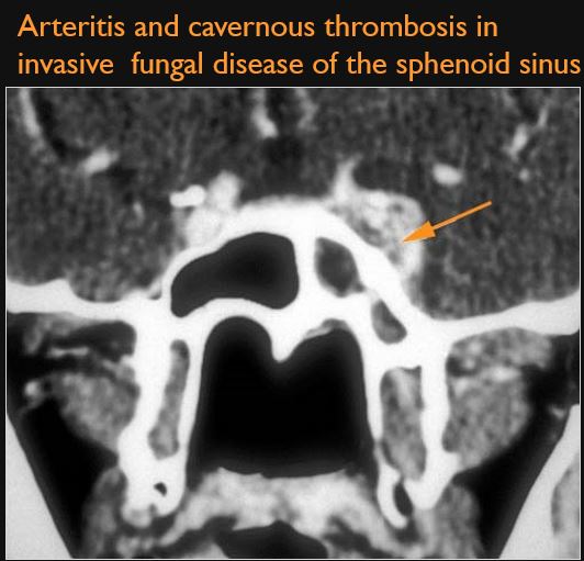
There is primary sinonasal, bone or skull base rather than a primary orbital disease process that might be causative and producing the patient’s signs and/or symptoms related to the orbit. [Yes/No]
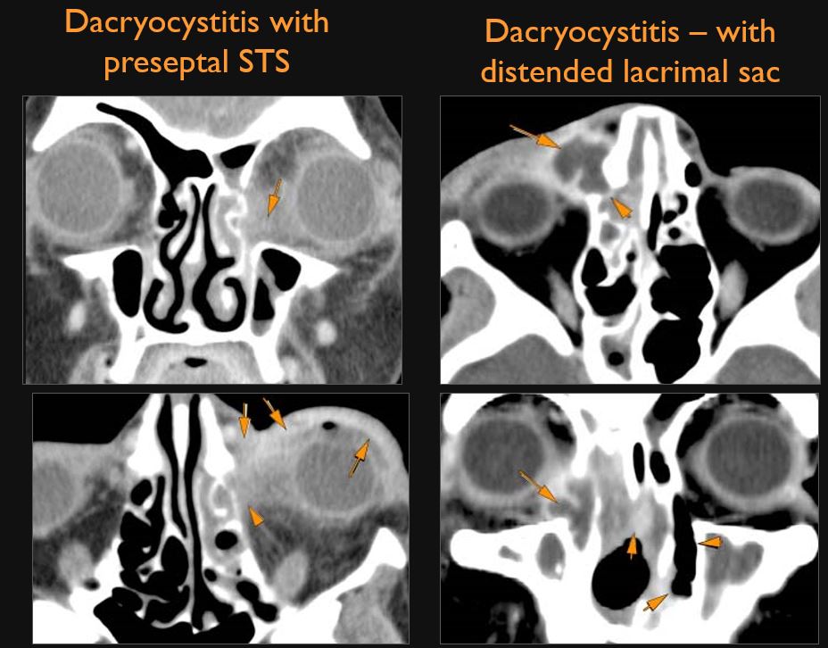
The pre-septal soft tissues including the lacrimal sac are abnormal. [Yes/No]
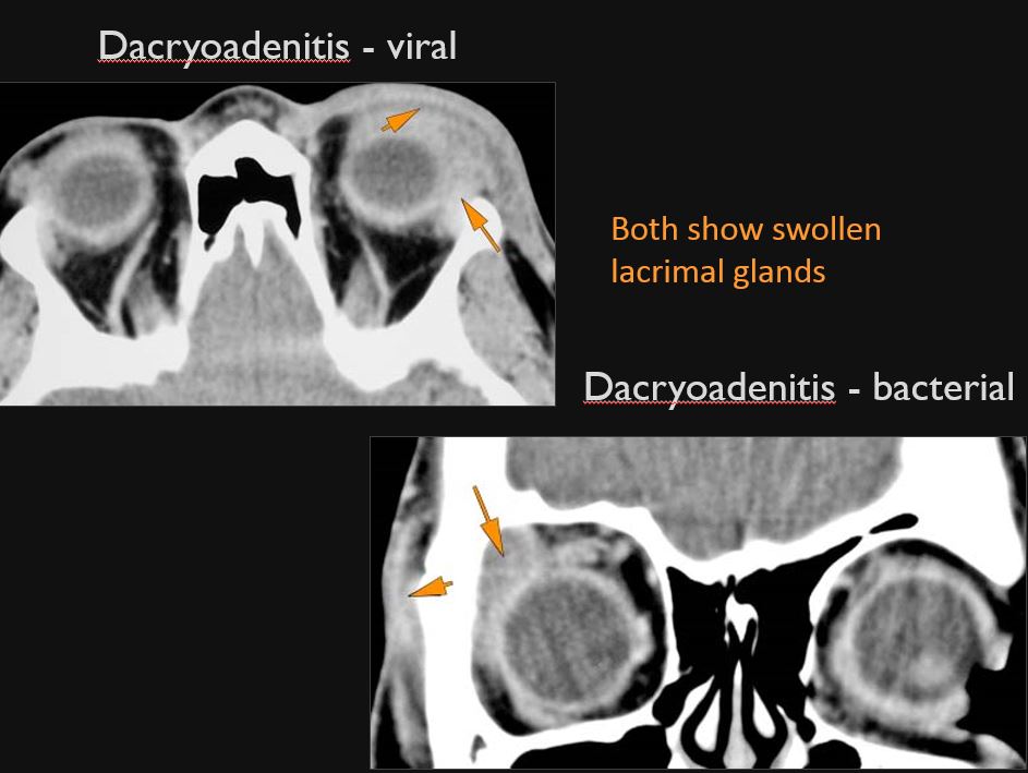
The lacrimal gland is abnormal. [Yes/No]
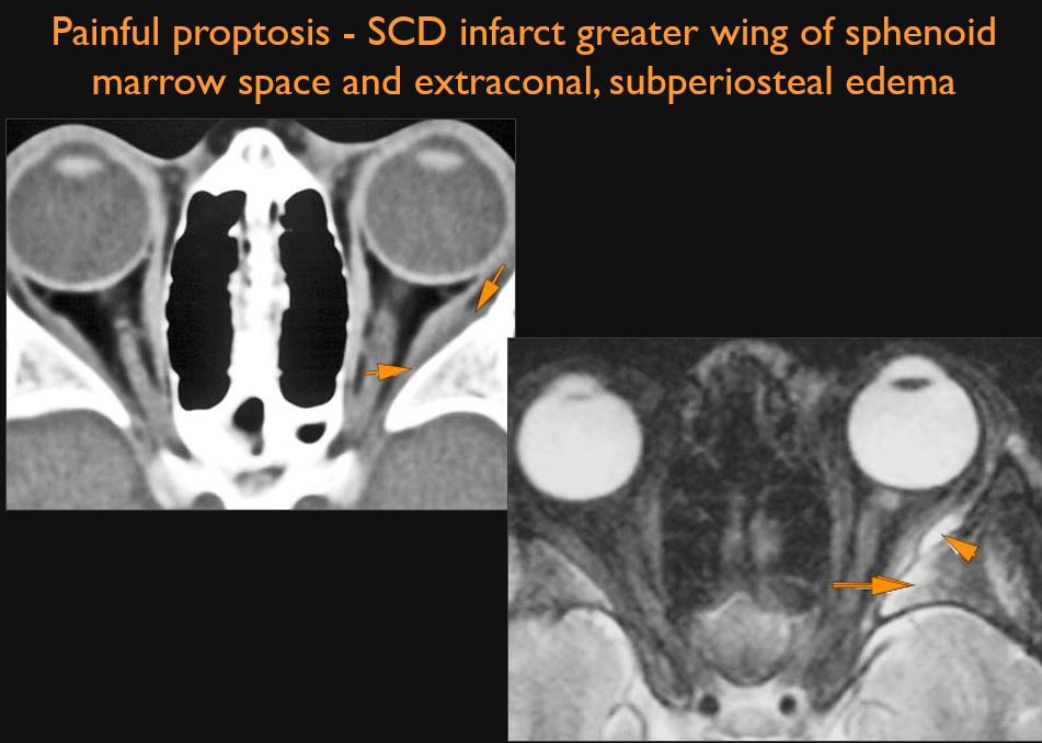
The bones and bony margins of the orbit including all component parts of the sphenoid bone are abnormal. [Yes/No]
There is a structural abnormality or infiltrating process of the extraconal or intraconal compartments of the orbit. [Yes/No]
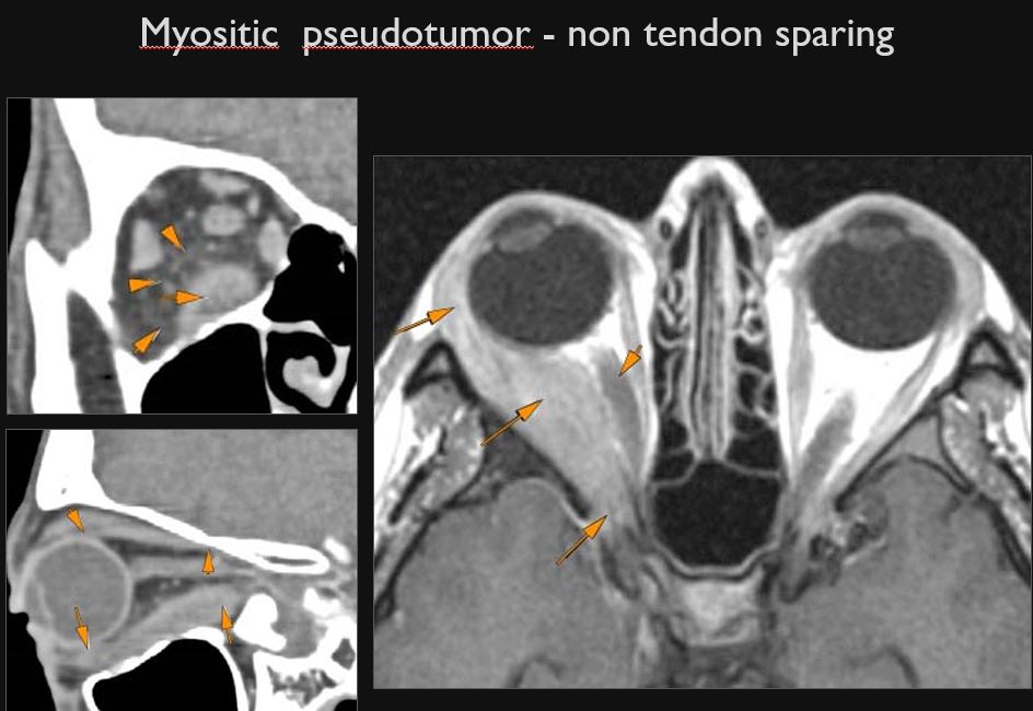
The extraocular muscles appear to be swollen or otherwise abnormal. [Yes/No]
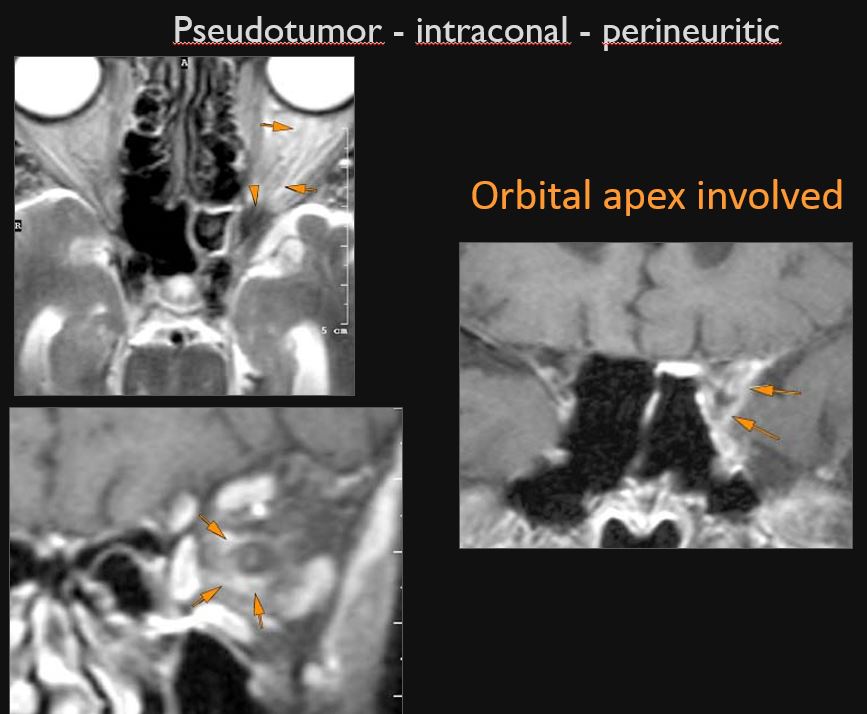
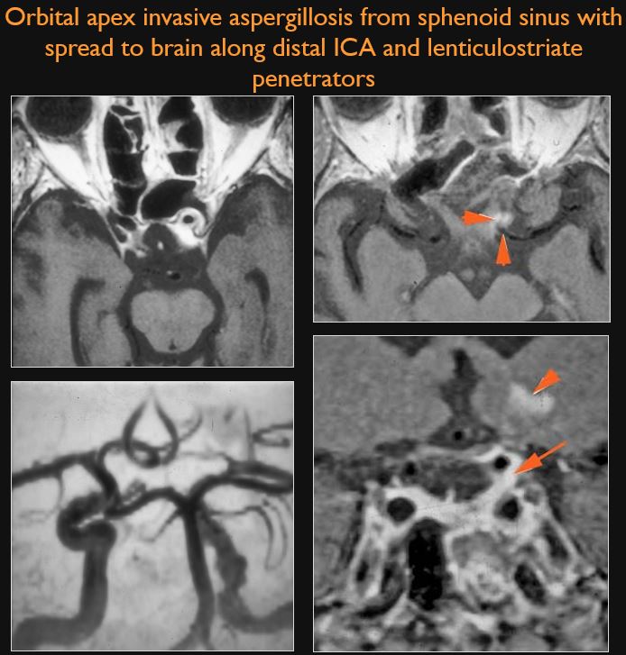
The orbital apex and superior orbital fissure are infiltrated, edematous or otherwise abnormal. [Yes/No]
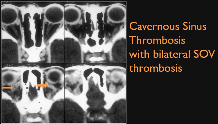
There is enlargement and/or thrombosis of the superior, inferior or other orbital veins. [Yes/No]
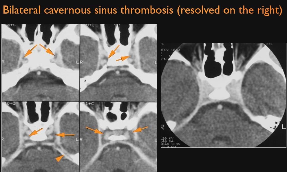
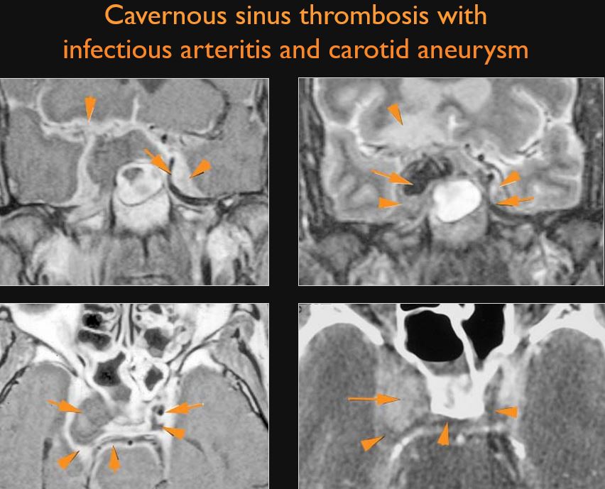
There is evidence of cavernous sinus thrombosis and/or inflammatory morphologic features in the cavernous sinus or para-cavernous region. [Yes/No]
Eye
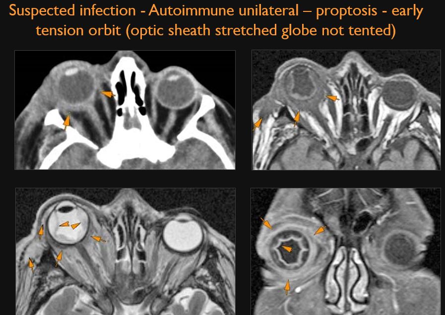
Proptosis is present. [Yes/No]
The optic nerve is stretched in appearance. [Yes/No]
The posterior aspect of the globe is tented in appearance. [Yes/No]
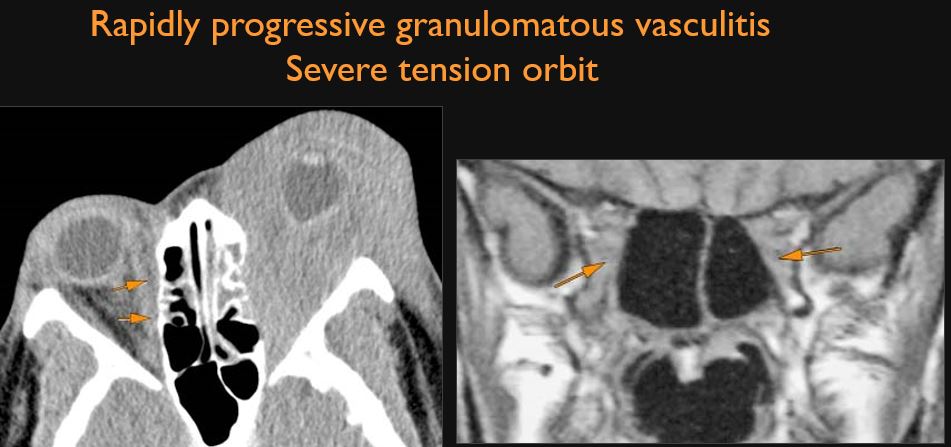
Signs of tension orbit are present. [Yes/No]
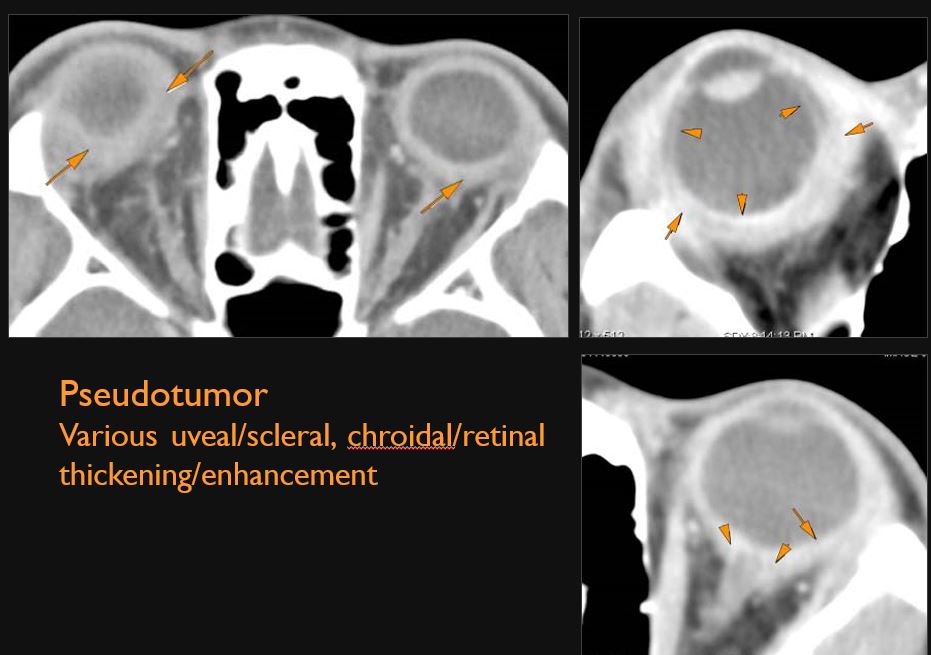
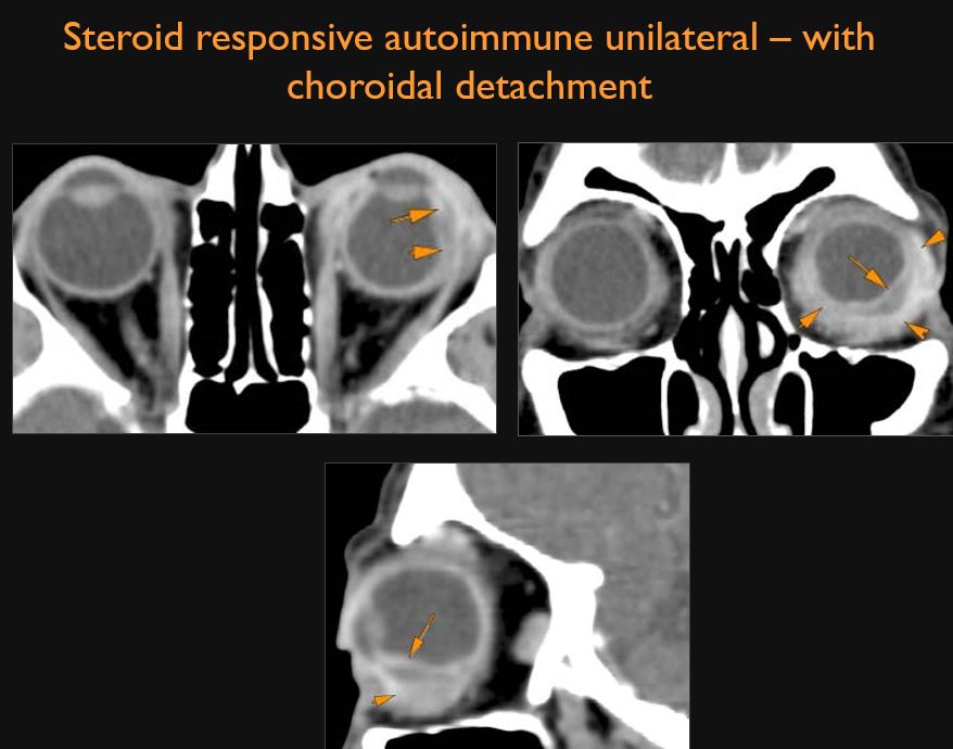
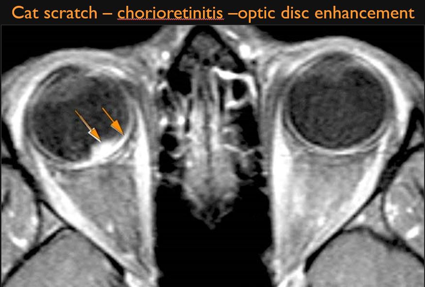
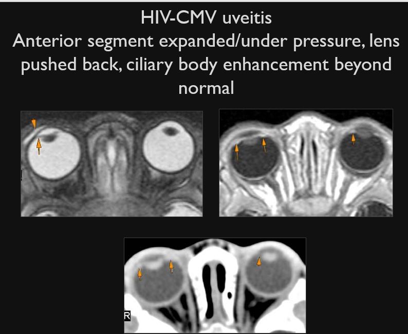
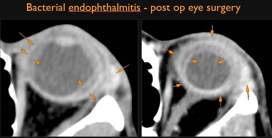
The uveal and scleral components of the globe are swollen or enhancing abnormally. [Yes/No]
There is a hemorrhage or other abnormality causing a detachment of the choroid, hyaloid membrane and/or retina. [Yes/No]
The optic disc region is abnormally swollen or enhancing. [Yes/No]
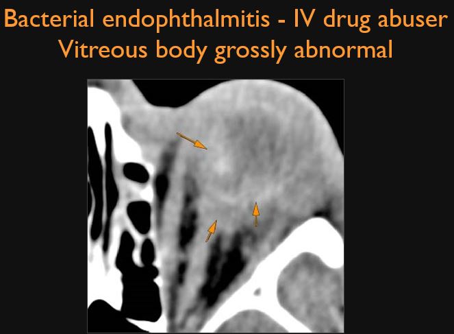
The vitreous body is abnormal in appearance. [Yes/No]
The anterior segment structures including the lens are abnormal in appearance. [Yes/No]
Signs of endophthalmitis are present. [Yes/No]
Optic nerve/sheath and chiasm
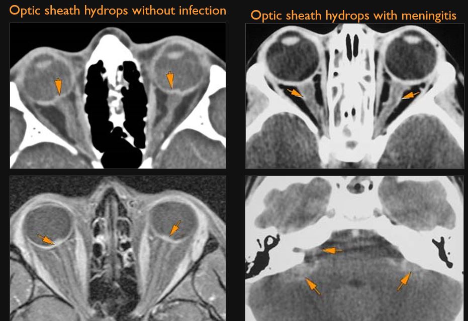
The optic nerves and/or optic sheaths appear abnormal in size. [Yes/No]
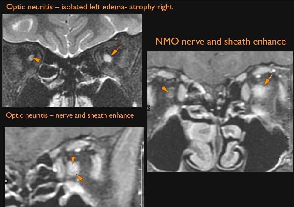
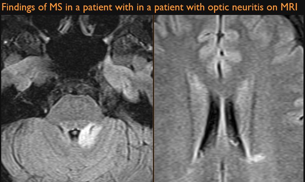
The optic nerves appear otherwise abnormal. [Yes/No]
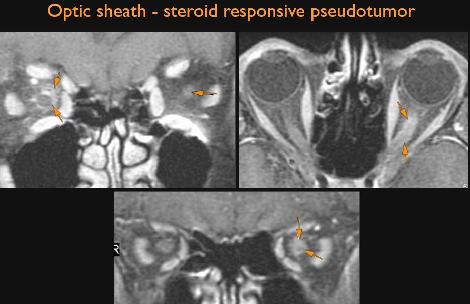
There is abnormal enhancement of the optic nerves and/or of the optic sheath. [Yes/No]
There is a structural abnormality or infiltrating process (especially around the optic sheath) of the intraconal compartment of the orbit. [Yes/No]
There is a compressive lesion or infiltrative process present anywhere along the course of the optic nerves, optic chiasm or optic tracts before entering the brain. [Yes/No]
There is a compressive lesion or infiltrative process present in the cavernous sinus or para-cavernous area. [Yes/No]
There is evidence of an aneurysm or carotid cavernous fistula present. [Yes/No]
There is abnormal meningeal or other enhancement of the optic chiasm, optic nerves or other structures in the suprasellar and/or chiasmatic cistern. [Yes/No]
There is generalized abnormal meningeal enhancement. [Yes/No]
Brain
There are intra-axial or extra-axial abnormalities of the brain that might be related to the orbital pathology. [Yes/No]
There is evidence of obstructive or communicating hydrocephalus. [Yes/No]
Impression
Ocular and Orbital Inflammation
-
There are findings to confirm an orbital or periorbital inflammatory or infectious process.
-
Specify the most likely diagnosis and/or most reasonable differential diagnostic alternatives.
