Case Notes
History
62 yo male with AML and feverExam
Prior Study
multiple prior examsDicom
Findings
| Technique | Correct Answer | Your Answer |
|---|---|---|
|
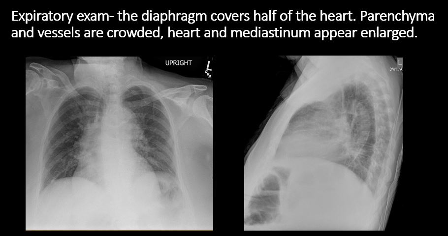
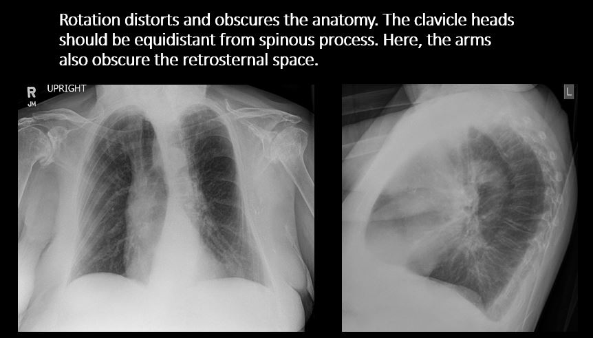
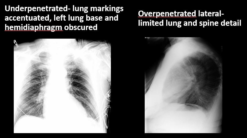
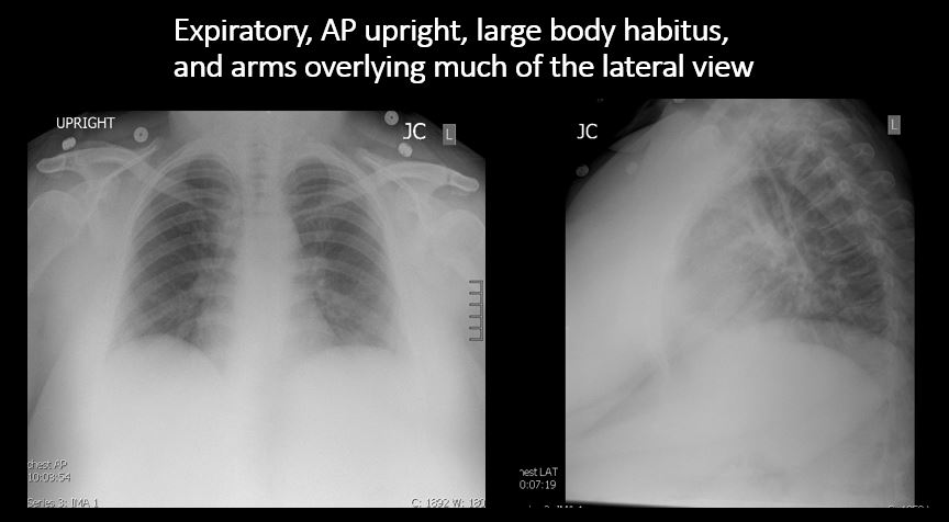
The exam is expiratory, rotated, over or under penetrated, or limited by overlying structures or soft tissues, body habitus, patient positioning, or motion. |
No | NA |
| Support Devices | Correct Answer | Your Answer |
|---|---|---|
|
There is a vascular line or lines present in an abnormal location or otherwise abnormal. |
Yes | NA |
|
There are other devices such as a chest tube or pleural drain, feeding, NG, or gastrostomy tube, LVAD or pacemaker, vascular stents, cardiac valves, anesthesia catheter, VP shunt, neurostimulator, or other drains or tubes present that are in an abnormal position or are otherwise abnormal. |
No | NA |
| Cardiomediastinum | Correct Answer | Your Answer |
|---|---|---|
|
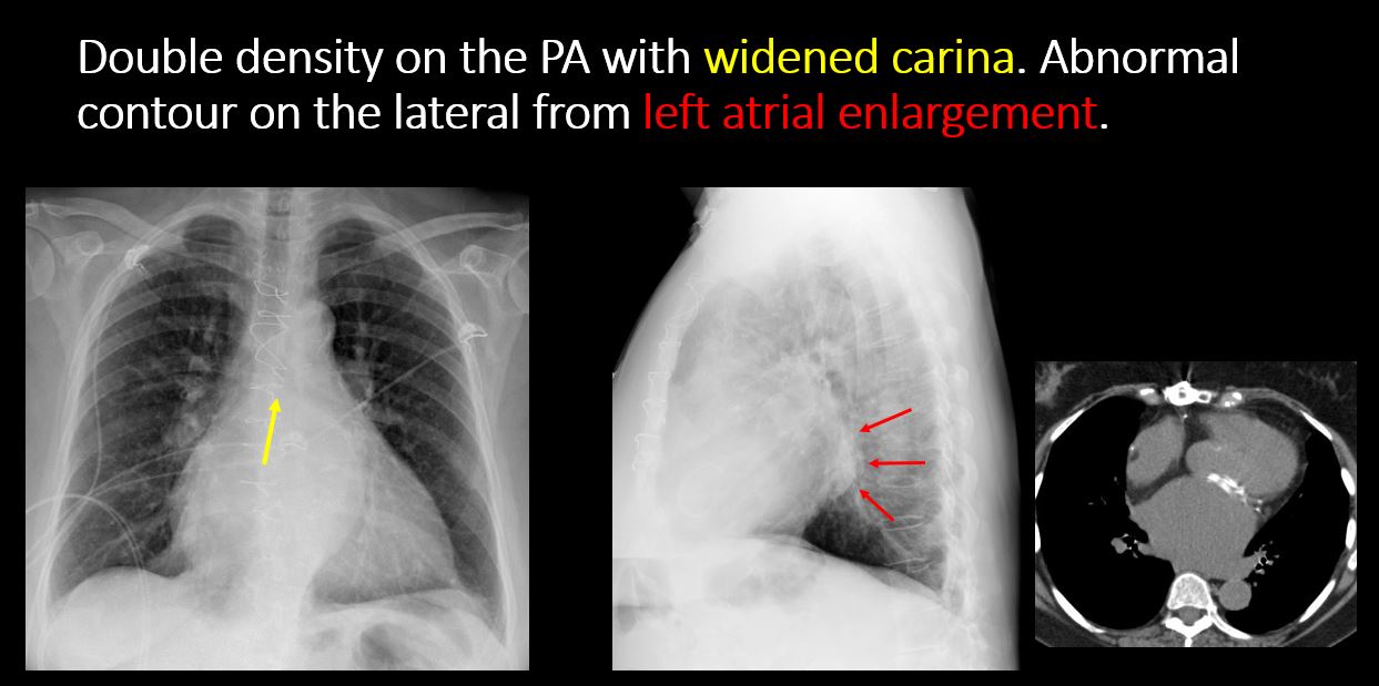
The superior mediastinum is abnormally widened considering the technique. |
No | NA |
|
There is abnormal shift of the mediastinum. |
No | NA |
|
There is a mass or other abnormal density in or overlying the mediastinum. |
Yes | NA |
|
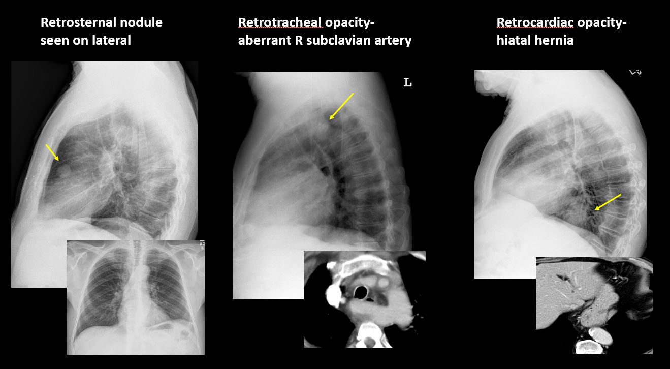
There is an abnormality in the retrosternal, retrotracheal, or retrocardiac space on the lateral view. |
Yes | NA |
|
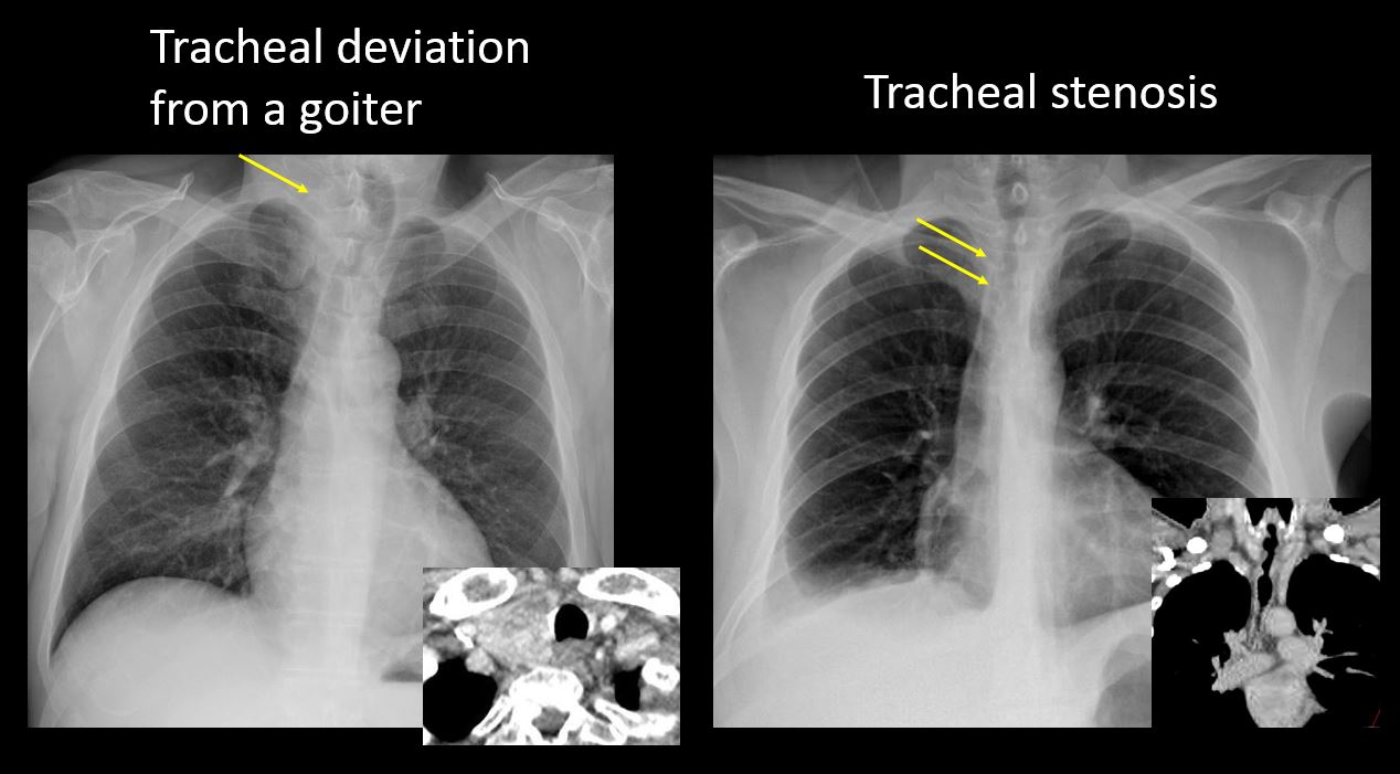
There is abnormal tracheal deviation or narrowing on the frontal and/or lateral view. |
No | NA |
|
|
No | NA |
|
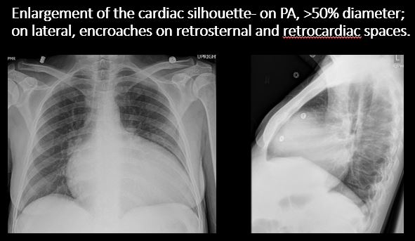
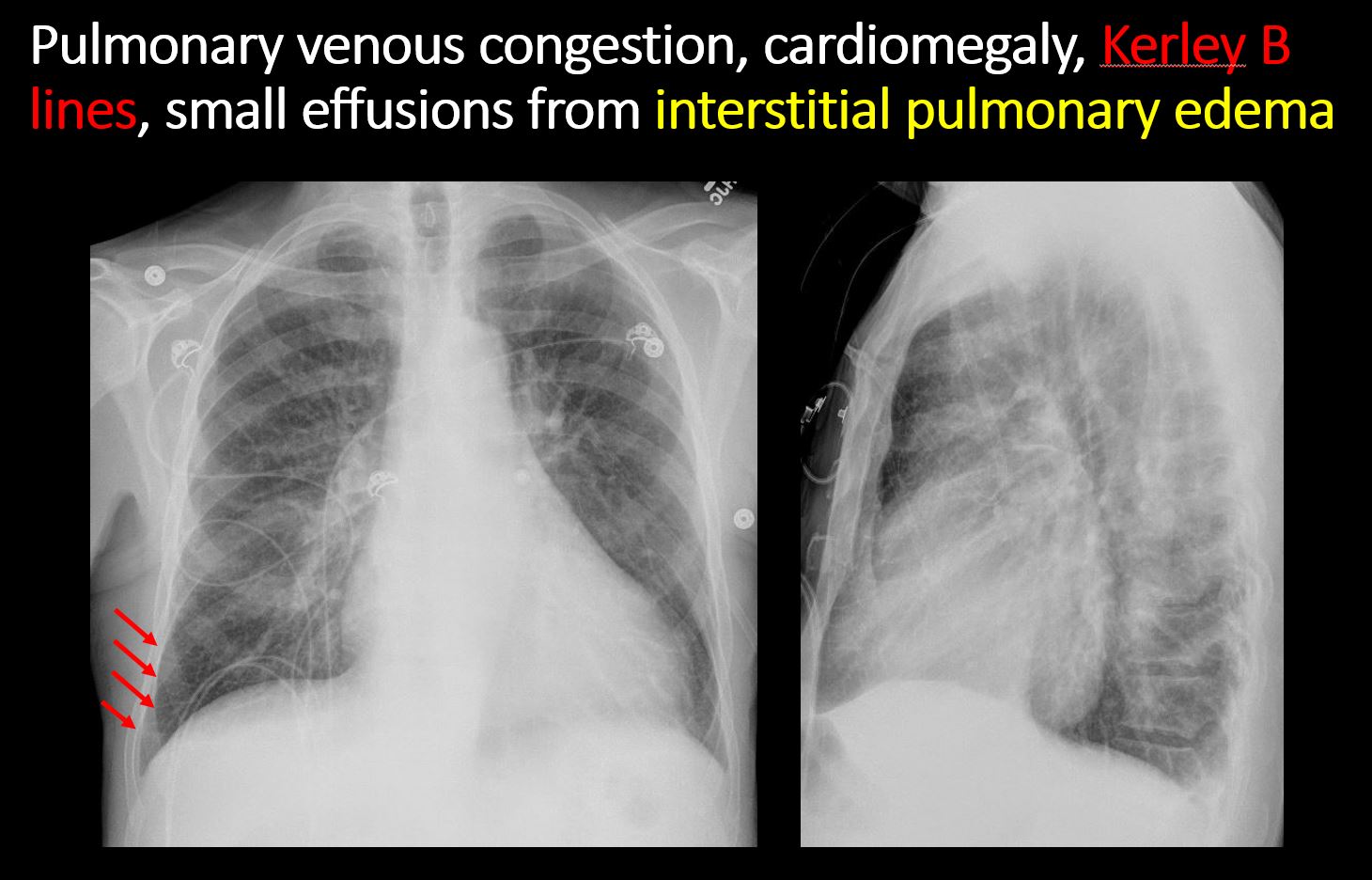
There is enlargement of the cardiac silhouette. |
No | NA |
|
|
No | NA |
|
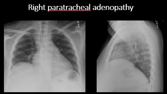
The right paratracheal stripe is thickened or enlarged. |
No | NA |
|
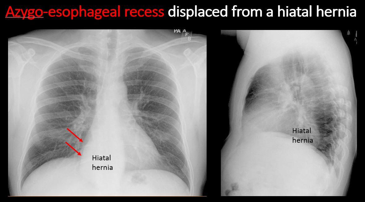
The azygoesophageal recess is displaced or otherwise obscured. |
Yes | NA |
| Vasculature and Hila | Correct Answer | Your Answer |
|---|---|---|
|
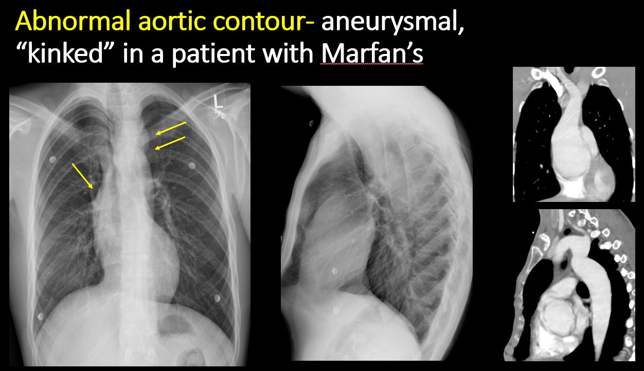
The aorta is dilated, tortuous, ectatic, calcified, or there is a focal contour abnormality. |
No | NA |
|
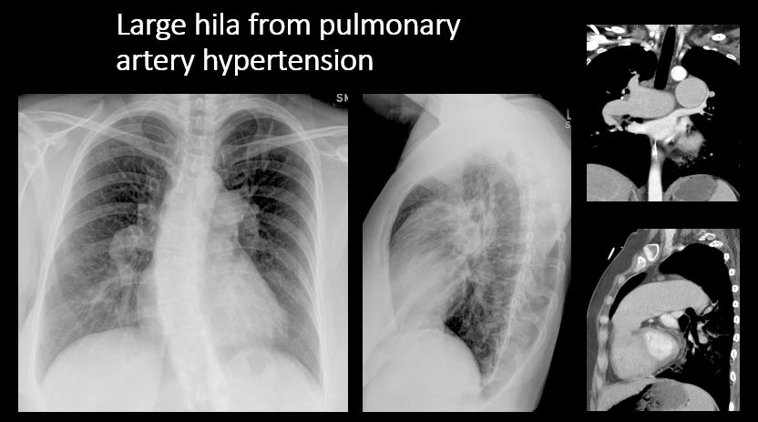
The central pulmonary arteries or hila are enlarged. |
No | NA |
|
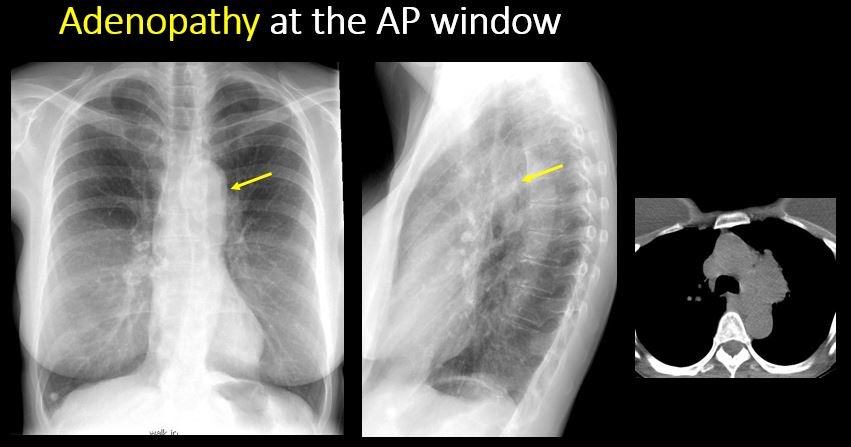
The aorto-pulmonary window is convex or obscured by a mass, adenopathy, or vasculature. |
No | NA |
|
|
No | NA |
| Lungs | Correct Answer | Your Answer |
|---|---|---|
|
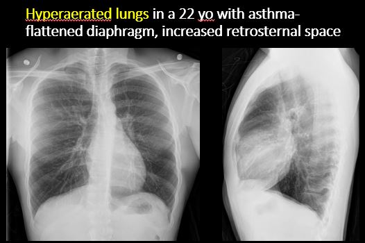
The lungs are hyperinflated or underinflated generally or segmentally. |
No | NA |
|
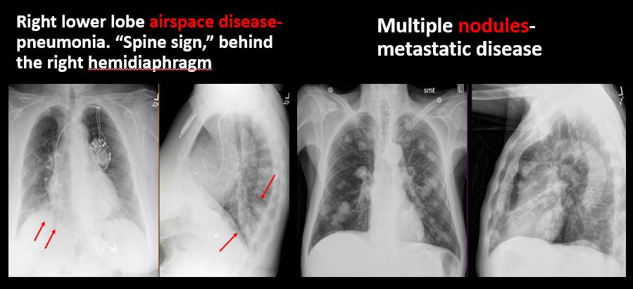
There is focal, multifocal, or diffuse airspace disease, mass, opacity, or nodularity. |
Yes | NA |
|
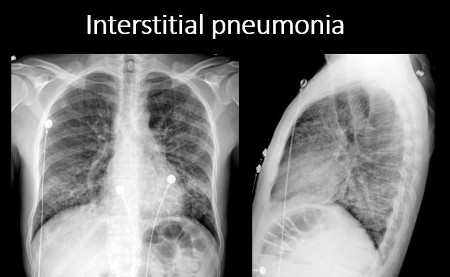
There is focal or diffuse interstitial disease. |
No | NA |
|
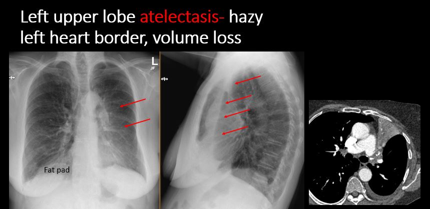
There is focal or lobar atelectasis or total collapse of the lung. |
Yes | NA |
|
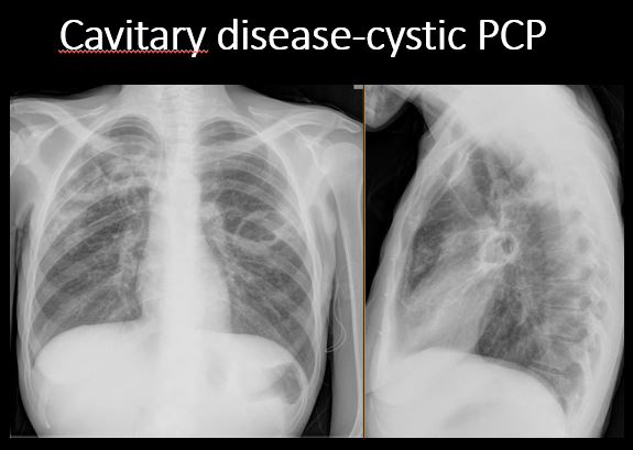
There is cavitary or cystic disease with or without air fluid levels or soft tissue nodularity. |
No | NA |
|
The position of the major and minor fissures is abnormal. |
Yes | NA |
|
The right or left hemidiaphragm is focally or diffusely obscured on the frontal and/or lateral view. |
Yes | NA |
|
There is elevation, depression, or contour abnormality of the right or left hemidiaphragm. |
No | NA |
| Pleura | Correct Answer | Your Answer |
|---|---|---|
|
There is focal or diffuse abnormality of the pleura or chest wall. |
No | NA |
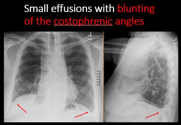
|
There is a pleural effusion, blunting of the costophrenic angle, or posterior sulci. |
Yes | NA |
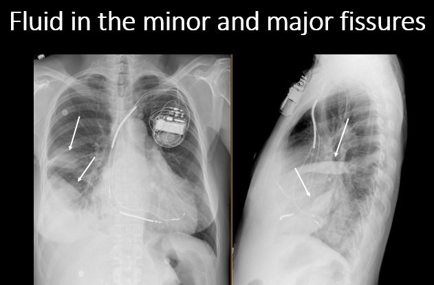
|
There is focal fluid in the fissures. |
Yes | NA |
|
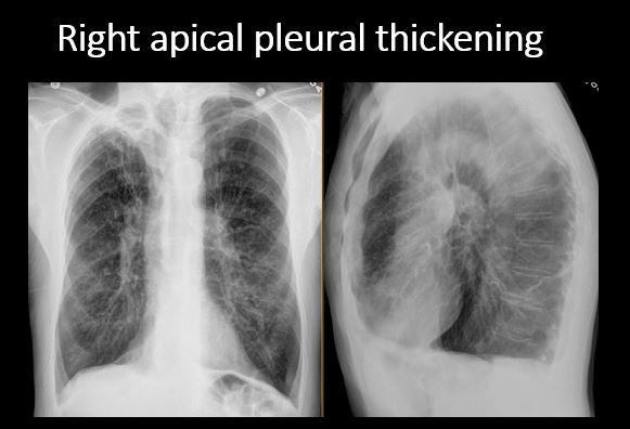
There is asymmetric pleural thickening or capping of the apices. |
No | NA |
|
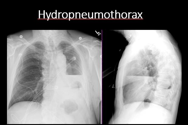
There is a pneumothorax, hydro/pneumothorax, or hemo/pneumothorax. |
No | NA |
| Bones, Soft Tissues, Upper Abdomen | Correct Answer | Your Answer |
|---|---|---|
|
The ribs, clavicles, shoulder, spine, or other visualized bones are abnormal. |
No | NA |
|
There is free air beneath the diaphragm. |
No | NA |
|
The bowel or organs of the upper abdomen are abnormal. |
No | NA |
|
There is subcutaneous emphysema, focal or diffuse soft tissue abnormality, radiopaque foreign body, or post-surgical change or hardware. |
No | NA |
Impression
Expert Answer
The left upper extremity PICC is in the azygous vein and needs to be retracted. There is a small to moderate right pleural effusion and parenchymal disease which could be from pneumonia in this patient with fever.
Your Answer
Recommendations & Acuity
Recommendations
Expert Answer
Verbally notify the clinical team about the malpositioned PICC in the azygous vein and need for retraction, and findings of pneumonia in this patient with fever.
Your Answer
Acuity
Expert Answer
Urgent (Action Necessary in a few hours)
