Case Notes
History
75 year old male with heart disease; PICC placementExam
Prior Study
Multiple prior portable chest exams.Dicom
Findings
| Technique | Correct Answer | Your Answer |
|---|---|---|
|
The exam is technically correct, and is not expiratory, is not rotated, nor over or under penetrated, nor limited by overlying structures or soft tissues, body habitus, patient positioning, or motion. |
yes | NA |
| Support Devices | Correct Answer | Your Answer |
|---|---|---|
|
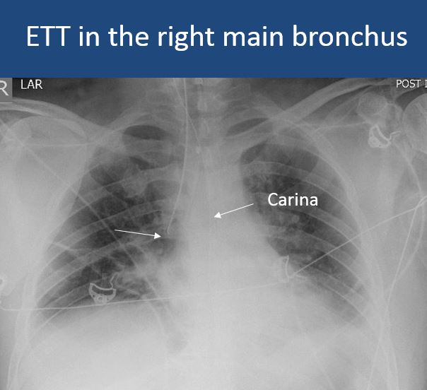
There is an ETT or tracheostomy tube presentand the ETT is less than 2cm from the carina or the ETT or tracheostomy tube is otherwise in abnormal position. |
no | NA |
|
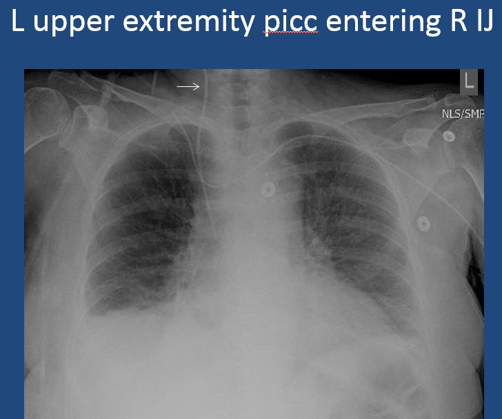
There is a venous line or lines present and in an improper location or other abnormality. |
no | NA |
|
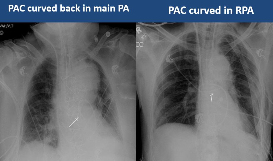
There is a PAC in place that is not in a main pulmonary artery or is otherwise abnormal. |
no | NA |
|
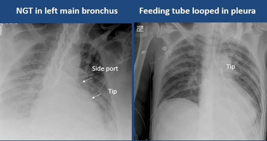
There is an NG tube and/or feeding tube in place and the NG or feeding tube does not go below the GE junction or has other abnormality. |
no | NA |
|
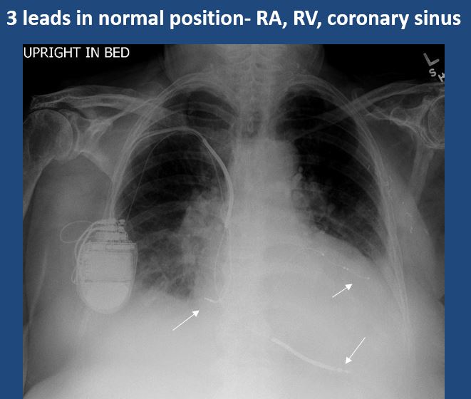
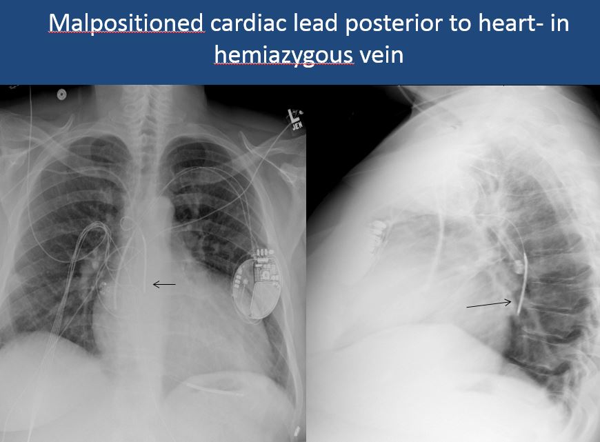
There is a cardiac pacemaker/AICD or temporary transvenous pacemaker the leads are fractured or in an abnormal location, not in the RA, RV, or coronary sinus. |
no | NA |
|
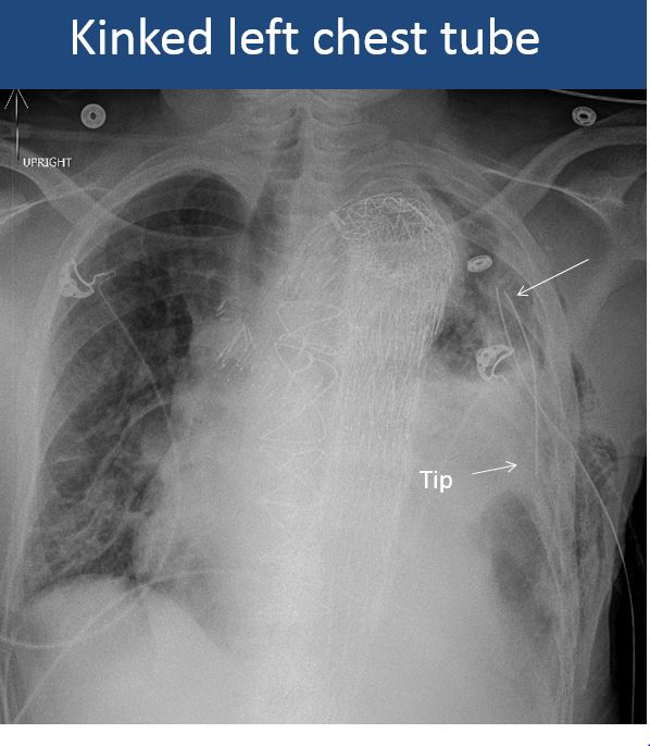
There is a chest tube in place and the side port of the chest tube is outside of the bony thorax or the tube is abnormally positioned. |
no | NA |
|
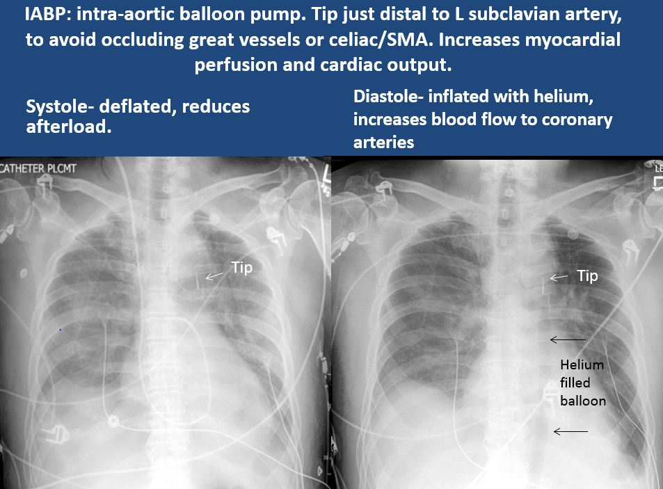
There are other devices such as an IABP, LVAD, vascular stents, cardiac valves, gastrostomy tube, other drains or tubes, anesthesia catheter, VP shunt, or neurostimulator present and improperly positioned. |
no | NA |
| Cardiomediastinum | Correct Answer | Your Answer |
|---|---|---|
|
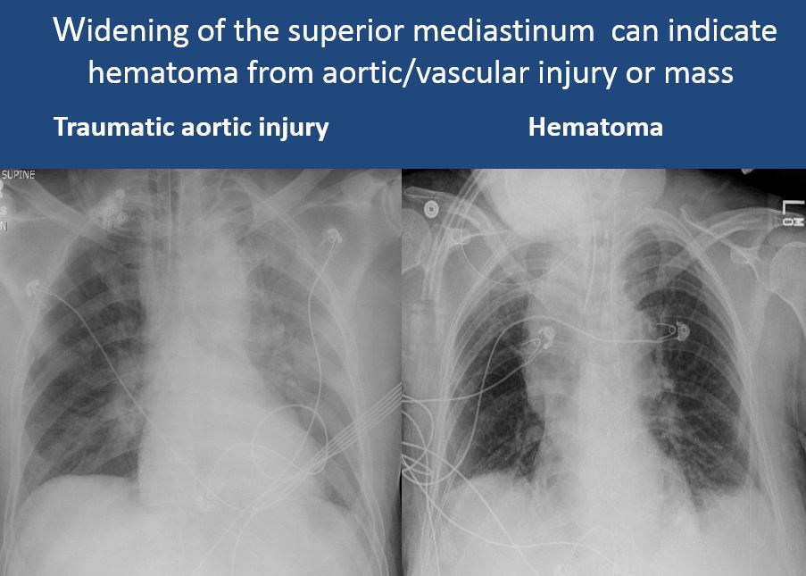
The mediastinum is abnormally widened considering the technique. |
No | NA |
|
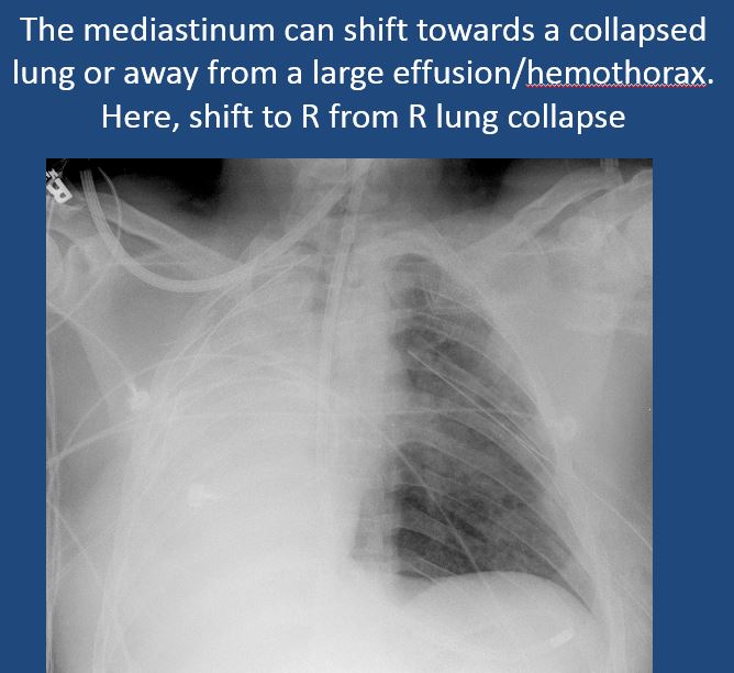
There is abnormal shift of the mediastinum. |
No | NA |
|
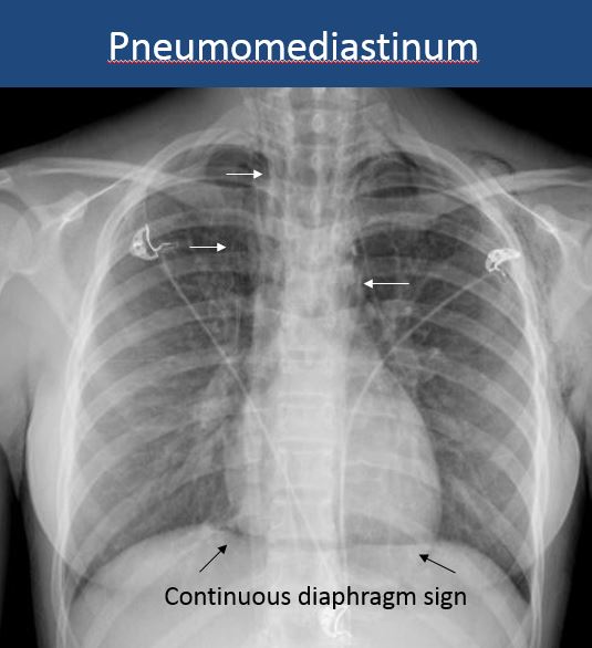
There is air in the soft tissues of the mediastinum. |
No | NA |
|
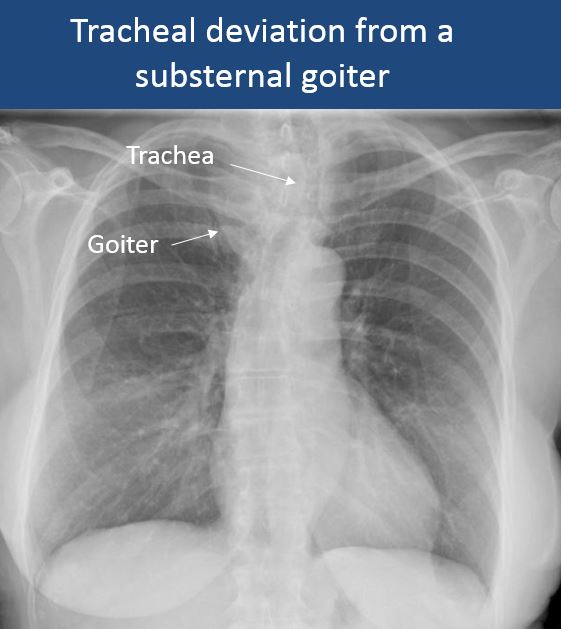
There is abnormal tracheal deviation or narrowing. |
No | NA |
|
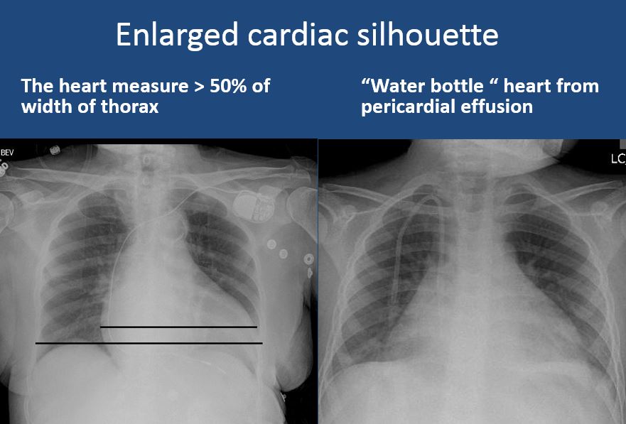
There is enlargement of the cardiac silhouette. |
Yes | NA |
|
|
No | NA |
|
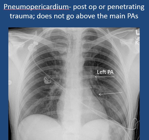
There is pneumopericardium. |
No | NA |
|
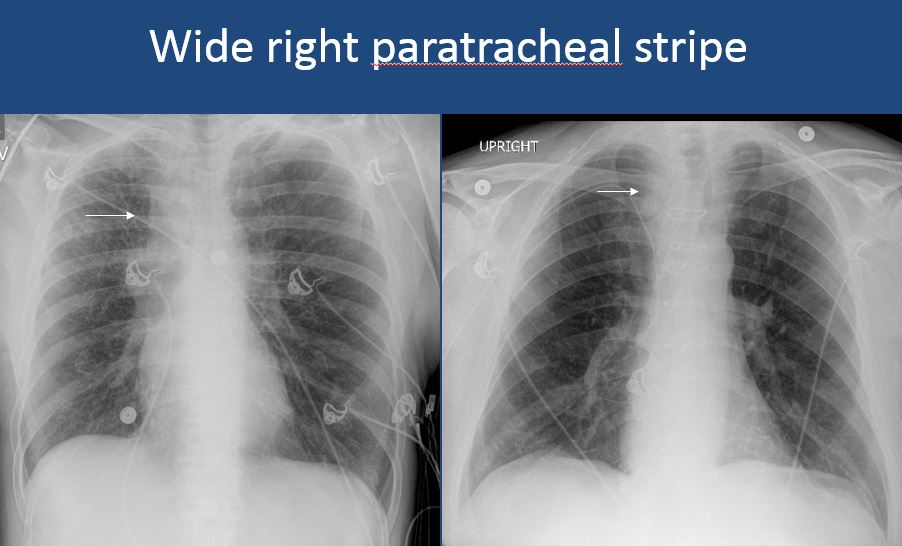
The right paratracheal stripe is thickened or enlarged. |
No | NA |
| Vasculature | Correct Answer | Your Answer |
|---|---|---|
|
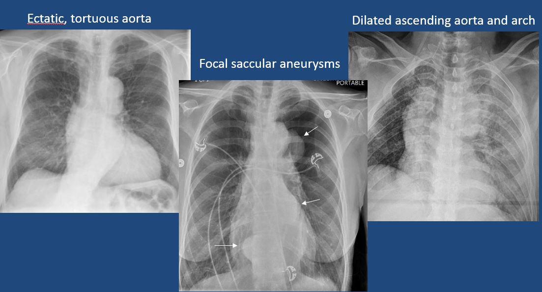
The aorta is dilated, tortuous, ectatic, calcified, or there is a focal contour abnormality. |
no | NA |
|
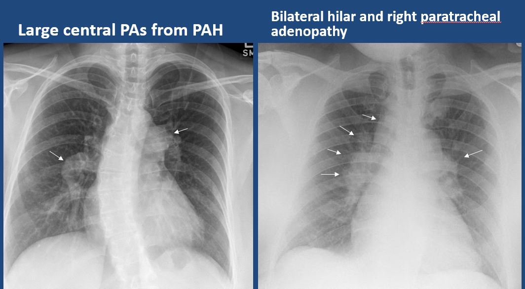
The central pulmonary arteries or hila are enlarged. |
No | NA |
|
|
Yes | NA |
| Lungs | Correct Answer | Your Answer |
|---|---|---|
|
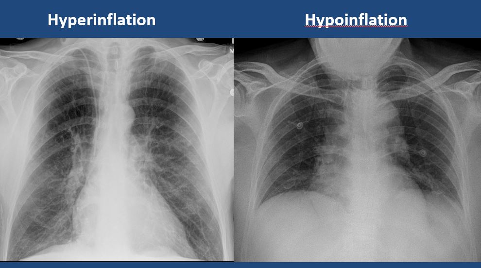
The lungs are hyperinflated or underinflated. |
no | NA |
|
|
yes | NA |
|
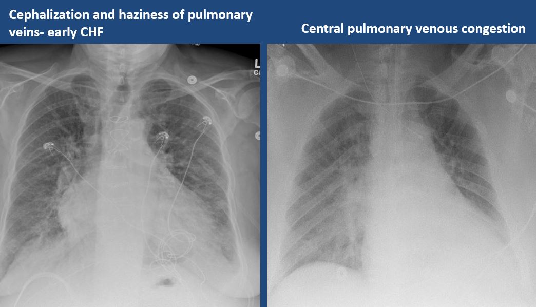
There is focal or diffuse interstitial disease. |
yes | NA |
|
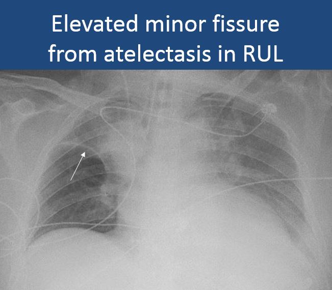
There is focal or lobar atelectasis or total collapse of the lung. |
yes | NA |
|
There is cavitary or cystic disease with or without air fluid levels or soft tissue nodularity. |
no | NA |
|
|
No | NA |
|
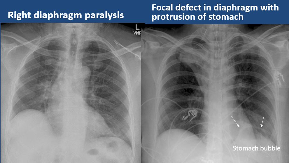
There is elevation, depression, or contour abnormality of the right or left hemidiaphragm. |
no | NA |
|
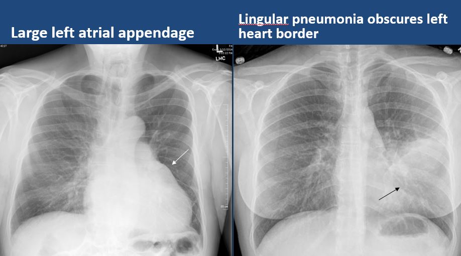
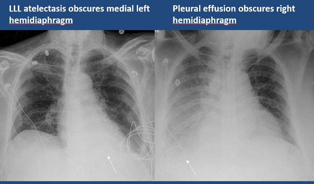
The right or left hemidiaphragm is focally or diffusely obscured. |
Yes | NA |
| Pleura | Correct Answer | Your Answer |
|---|---|---|
|
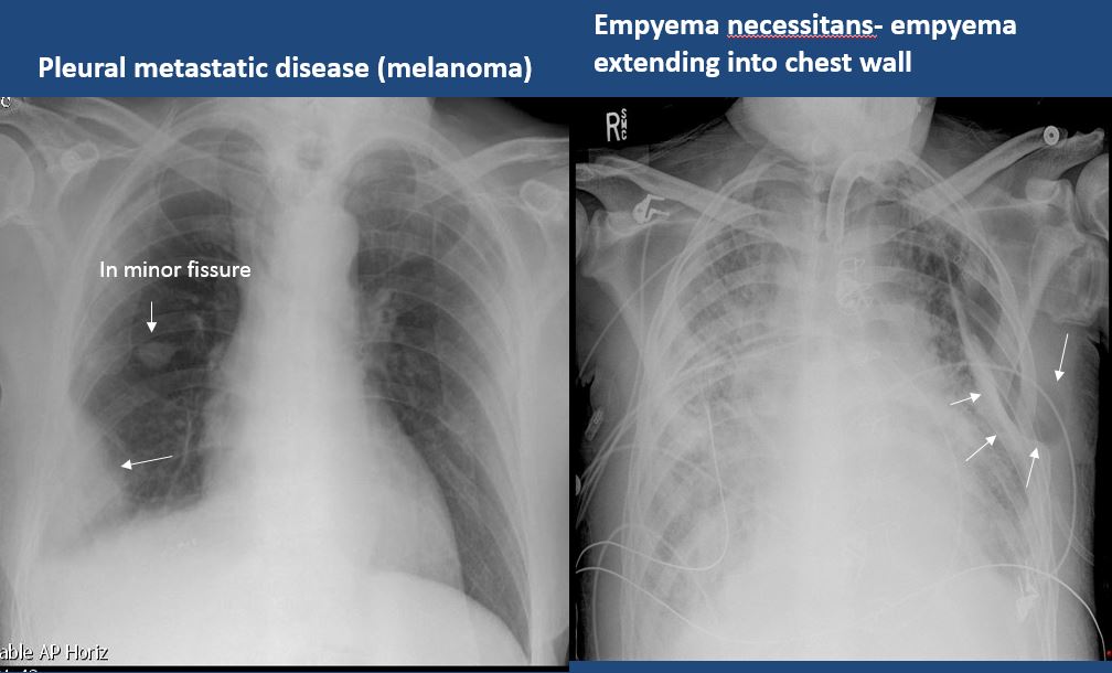
There is focal or diffuse abnormality of the pleura or chest wall. |
no | NA |
|
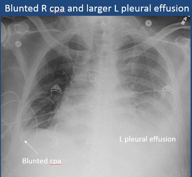
There is a pleural effusion or blunting of the costophrenic angle. |
Yes | NA |
|
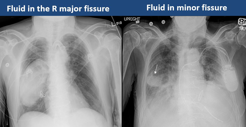
There is focal fluid in the fissures. |
Yes | NA |
|
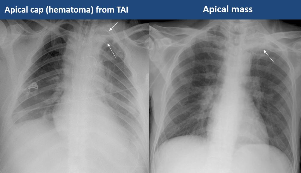
There is asymmetric pleural thickening or capping of the apices. |
No | NA |
|
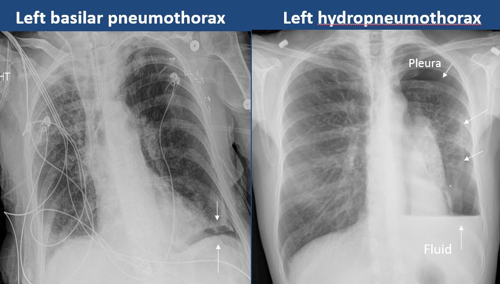
There is a pneumothorax, hydro/pneumothorax, or hemo/pneumothorax. |
no | NA |
| Bones, Soft Tissues, Upper Abdomen | Correct Answer | Your Answer |
|---|---|---|
|
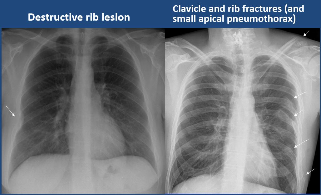
The ribs, clavicles, shoulder, spine, or other visualized bones are abnormal. |
no | NA |
|
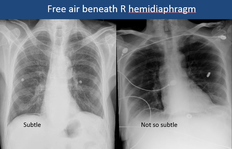
There is free air beneath the diaphragm. |
No | NA |
|
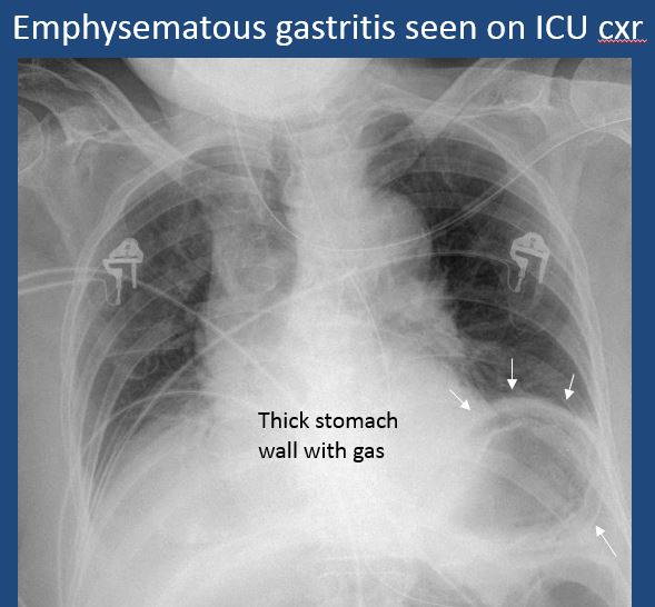
The bowel or organs of the upper abdomen are abnormal. |
no | NA |
|
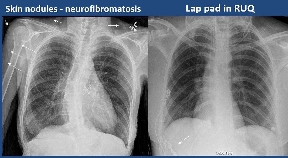
There is subcutaneous emphysema, focal or diffuse soft tissue abnormality, radiopaque foreign body, or post-surgical change or hardware. |
yes | NA |
