Case Notes
History
11 year old male with right hip pain.Exam
Prior Study
noneDicom
Findings
| Technique: | Correct Answer | Your Answer |
|---|---|---|
|
The exam is over or under penetrated. |
No | NA |
|
The exam is limited by overlying structures or soft tissues, body habitus, patient positioning, support devices, or motion. |
No | NA |
|
The area of concern is indicated by the patient, technologist, or care provider. |
Yes | NA |
|
The area of concern is included on the exam. |
Yes | NA |
| Soft Tissues: | Correct Answer | Your Answer |
|---|---|---|
|
There is soft tissue swelling, indistinctness of fat/muscle planes, gas, or laceration in the area of clinical concern. |
No | NA |
|
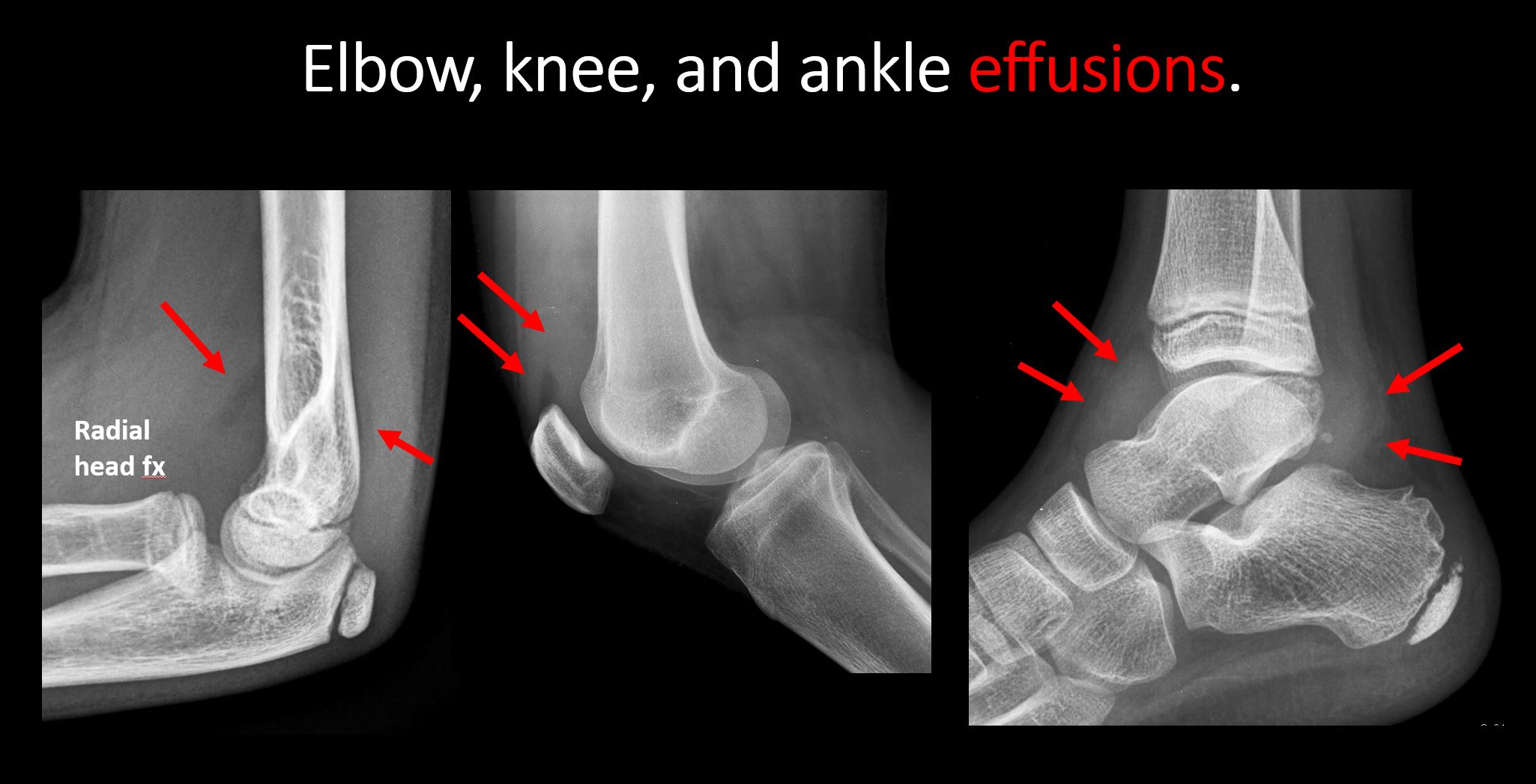
There is an effusion, fat pad displacement, or fat fluid level. |
No | NA |
|
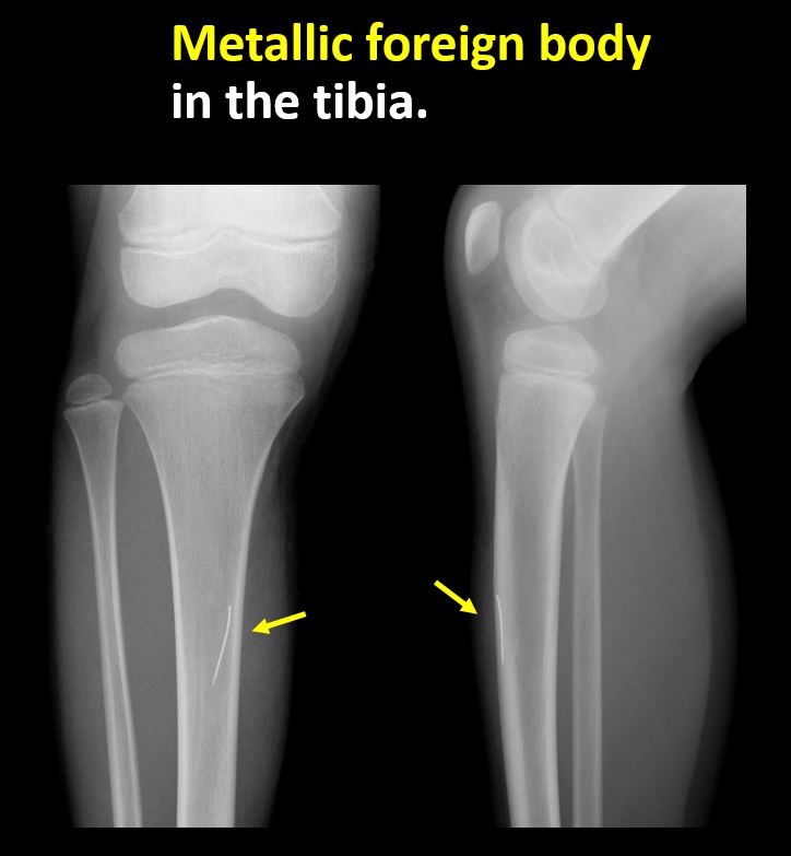
There is a radiodense or lucent foreign body. |
No | NA |
|
There are other densities, calcifications, post-surgical changes, or support devices in the soft tissues. |
No | NA |
| Bone: | Correct Answer | Your Answer |
|---|---|---|
|
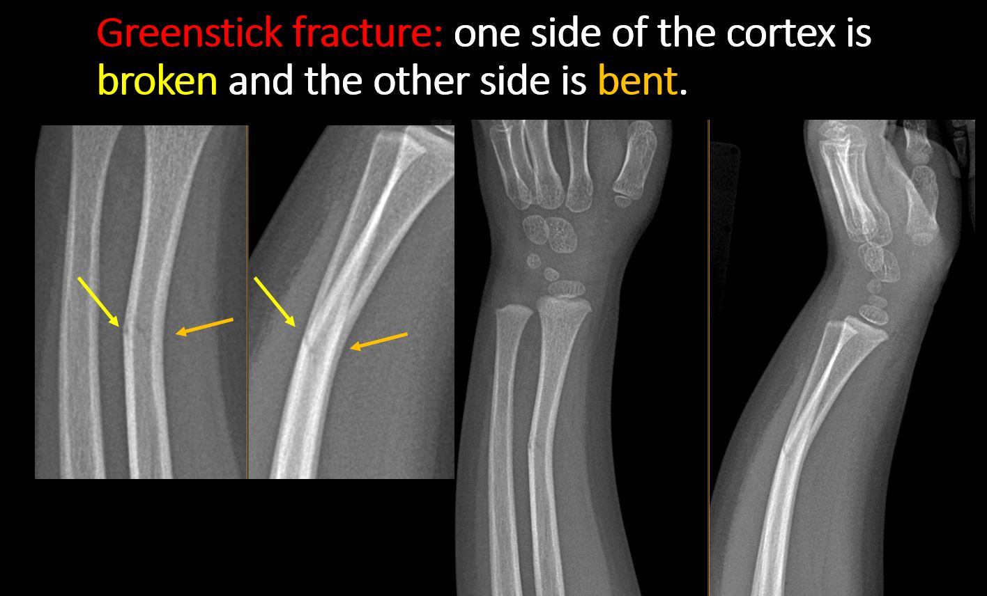
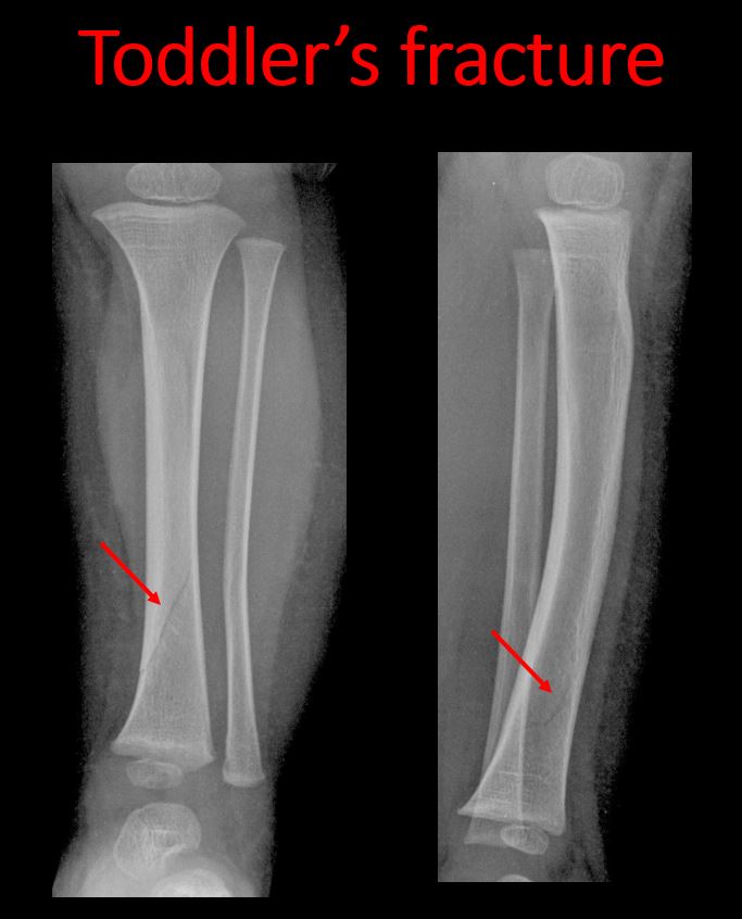
There is a break or interruption of the continuity of the cortical or cancellous bone with or without displacement of a fracture fragment which could be from a greenstick or toddler’s fracture, or other fracture. |
No | NA |
|
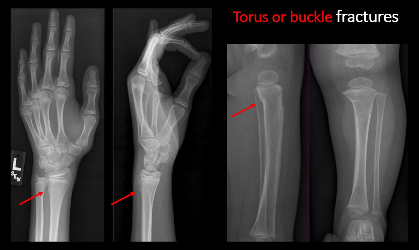
There is abnormal angulation or bulging of the cortical surface relative to the normal cortex which could be from a buckle or torus fracture. |
No | NA |
|
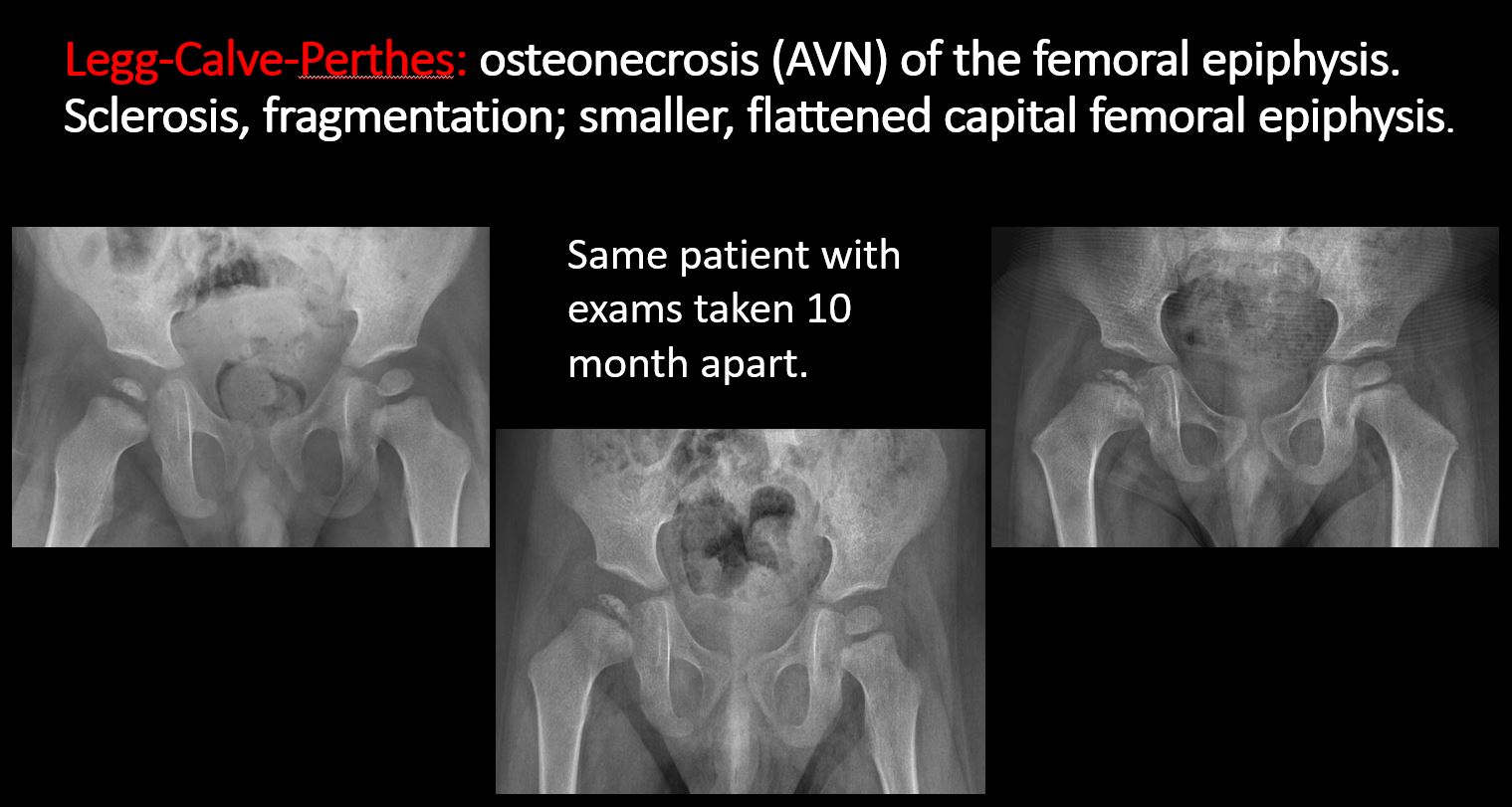
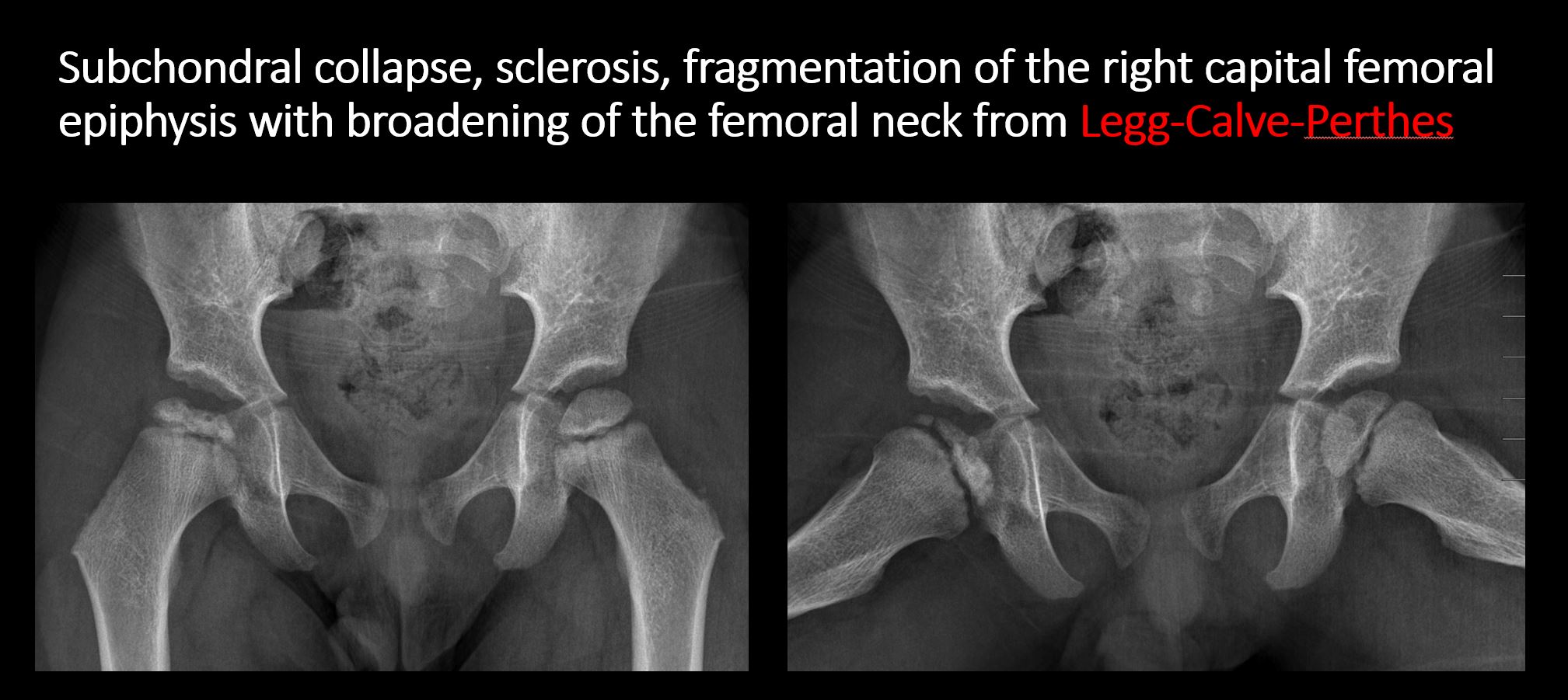
There is linear or irregular lucency, or increased density, cortical depression, flattening, or collapse, with or without cortical disruption or thickening, which may be from a compression or impaction fracture, stress or insufficiency fracture, osteonecrosis (e.g. Legg-Calve-Perthes), other fracture, or from growth recovery lines. |
No | NA |
|
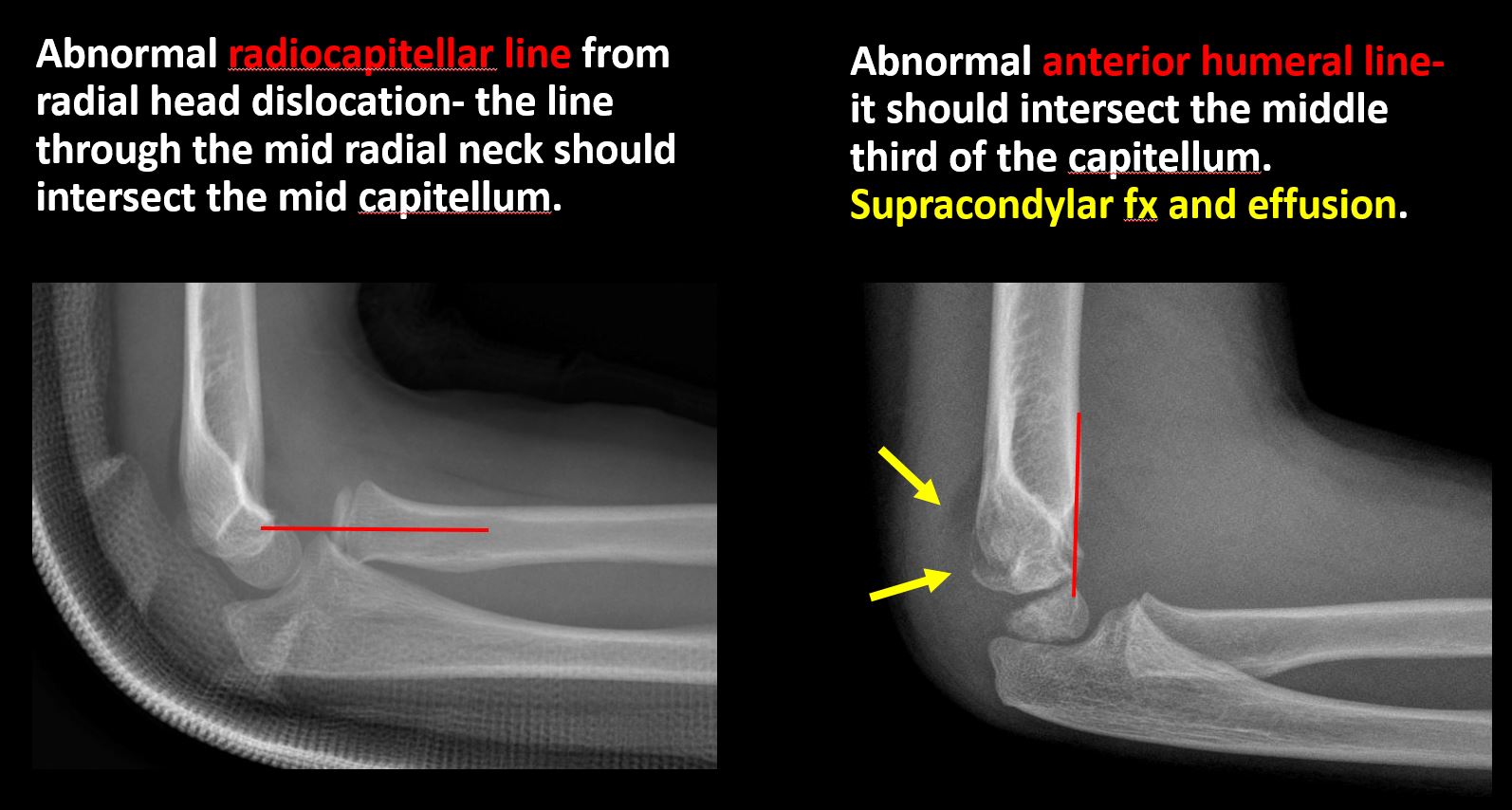
There is disruption of commonly recognized anatomical lines (e.g. iliopectineal, radiocapitellar, anterior humeral) or structures (e.g. sacral foraminal or arcuate lines). |
No | NA |
|
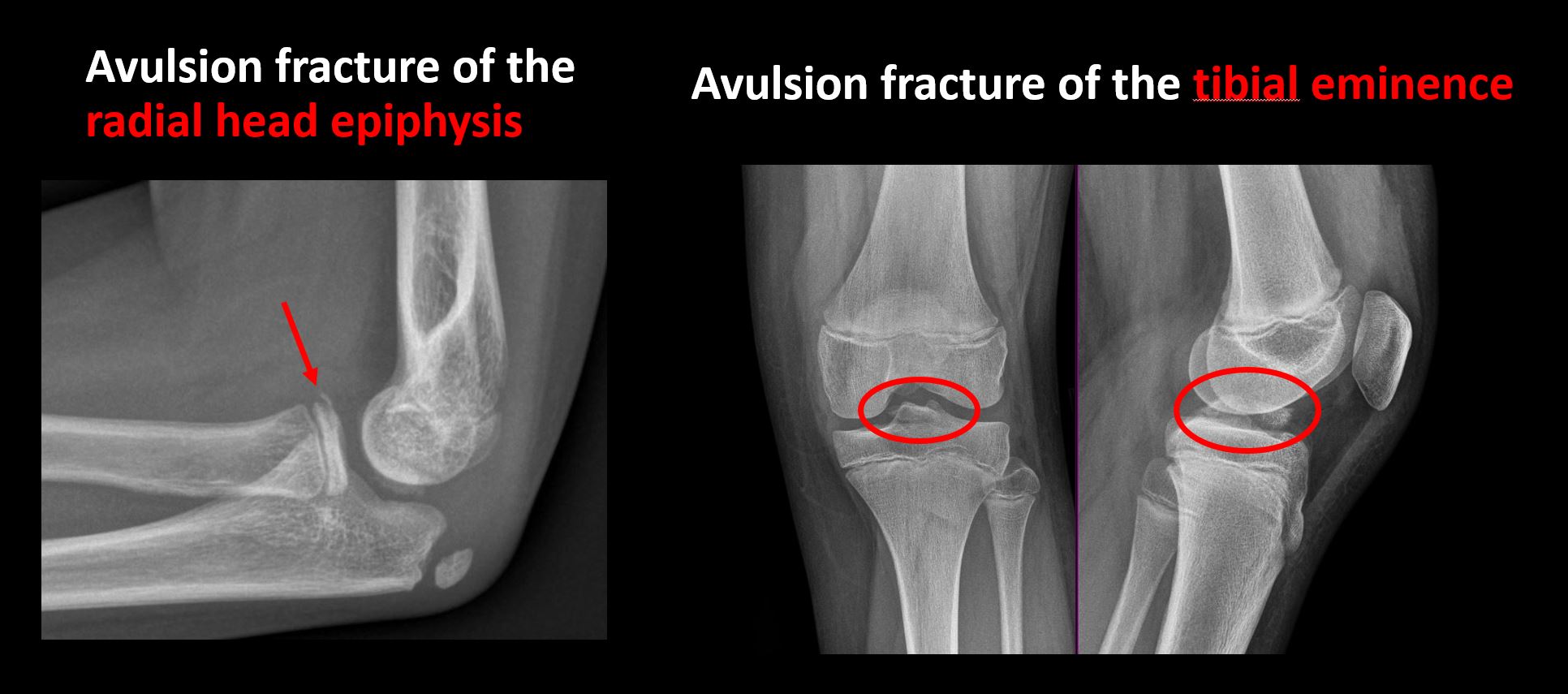
There is a displaced fragment which may be from avulsion by a tendon, ligament, or joint capsule or from a comminuted or other fracture. |
No | NA |
|
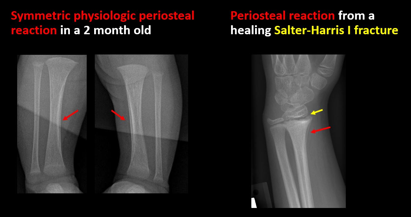
There is periosteal or endosteal reaction which could indicate a healing or subacute fracture, infection/inflammation, or other abnormality. |
No | NA |
|
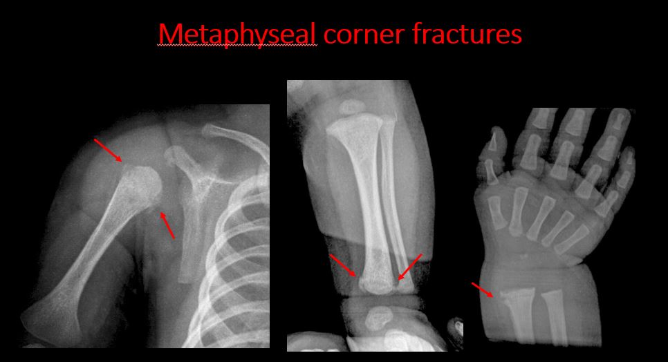
There is a corner fracture or metaphyseal lesion that could be from nonaccidental trauma. |
No | NA |
|
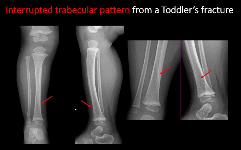
The stress trabeculae or other trabeculae of the cancellous bone are interrupted or otherwise abnormal. |
No | NA |
|
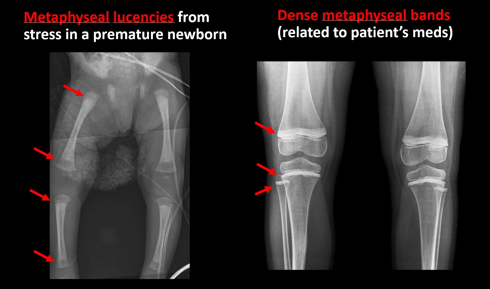
There is metaphyseal abnormality (lucencies, increased density, erosion) which may be from something other than injury such as stress, metabolic disease (e.g. rickets), neoplasm (e.g. leukemia), heavy metals, inflammation, or infection. |
No | NA |
|
There is/are focal or multifocal lytic/lucent, blastic/sclerotic or mixed density lesion(s) or other abnormality. |
No | NA |
|
Overall bone density is increased or decreased with or without thinning or thickening of the cortical or cancellous bone. |
No | NA |
| Growth plates, ossification centers, apophyses: | Correct Answer | Your Answer |
|---|---|---|
|
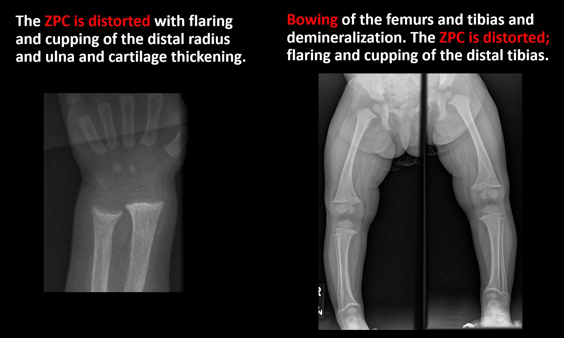
The growth plate(s) is/are abnormal. |
Yes | NA |
|
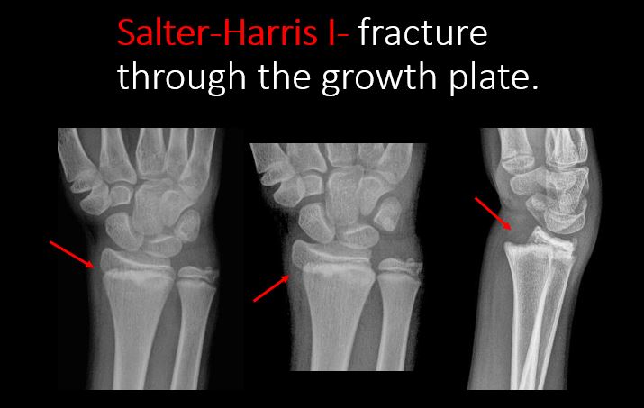
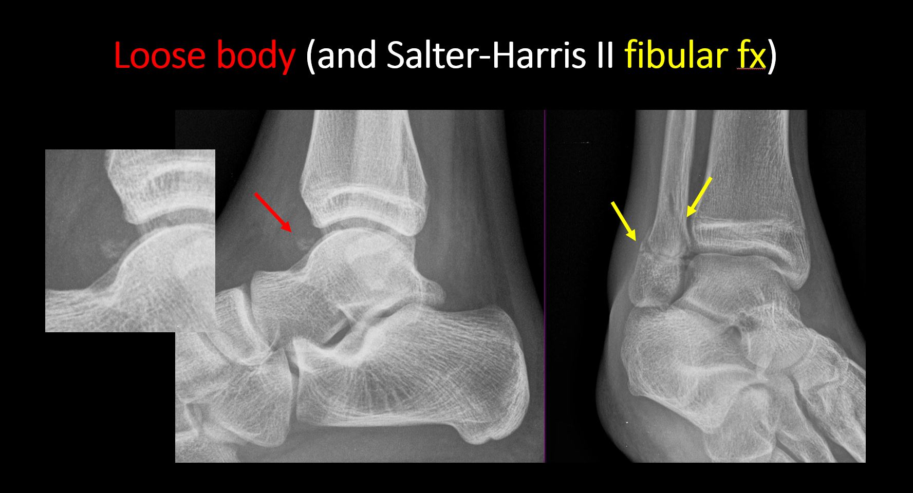
There is widening of the physis from a fracture with or without displacement of the epiphysis (Salter-Harris I). |
Yes | NA |
|
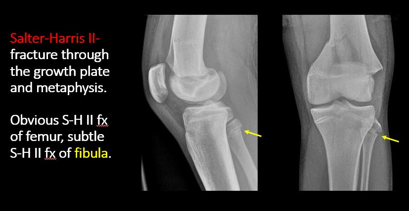
There is a fracture through the physis which then extends into the metaphysis with or without angulation or displacement (S-H II). |
N/A | NA |
|
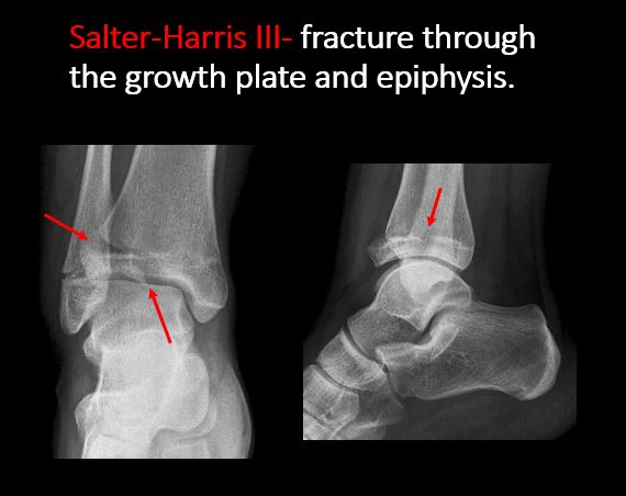
There is a fracture through the physis which then extends into the epiphysis and is intra-articular, with or without angulation or displacement (S-H III). |
N/A | NA |
|
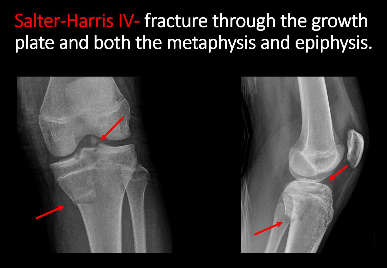
There is a fracture through the metaphysis, physis, and epiphysis which extends into the joint space with or without angulation or displacement (S-H IV). |
N/A | NA |
|
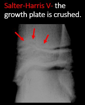
There is narrowing of the physis from a compression fracture (S-H V). |
N/A | NA |
|
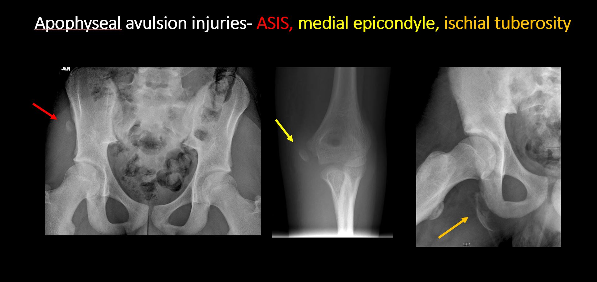
The apophysis, epicondyle, secondary ossification center, or accessory ossicle is displaced or otherwise abnormal (e.g. avulsed ASIS, medial epicondyle of the humerus, etc). |
No | NA |
| Joints and alignment: | Correct Answer | Your Answer |
|---|---|---|
|
There is an effusion, fat pad displacement, or fat fluid level. |
No | NA |
|
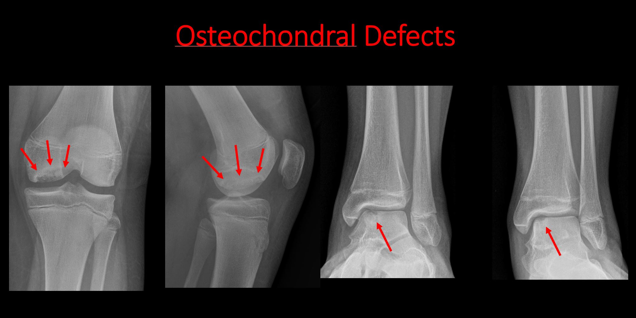
The epiphysis or subchondral bone is fractured, interrupted, flattened, compressed, impacted, displaced, or otherwise abnormal. |
Yes | NA |
|
There is an intra-articular loose body or chondrocalcinosis. |
No | NA |
|
The joint is widened, narrowed, dislocated, malaligned, or incongruent. |
No | NA |
|
There is pseudoarthrosis. |
No | NA |
| Other findings: | Correct Answer | Your Answer |
|---|---|---|
|
There are developmental changes or other anatomic variants or other existing conditions that may or may not be contributing to symptoms which can or should be further evaluated non-emergently or are otherwise incidental. |
No | NA |
|
The remainder of the exam is abnormal for age. |
No | NA |
Impression
Expert Answer
There is a right slipped capital femoral epiphysis with mild posterior inferior displacement of the femoral head epiphysis (Salter-Harris I fracture).
Your Answer
Recommendations & Acuity
Recommendations
Expert Answer
Verbal communication of a slipped capital femoral epiphysis (SCFE) of the right hip.
Your Answer
Acuity
Expert Answer
Urgent (Action Necessary in a few hours)
