Retake
V4) Facial and upper extremity swelling
Review the Learning Outcomes, Hx, PE and Labs, and begin the module with your Provisional Diagnosis. Keep hitting "Next" to move through the module.
Learning Outcomes
- Articulate your relationship with the consulting diagnostic radiologists in the evaluation of a patient with generalized swelling.
- Review the DDx considerations in a patient with generalized swelling.
- Identify the spectrum of imaging findings in appropriate modalities for evaluating a patient with generalized swelling.
History
A 60-year-old female with an 80-pack year smoking history presents due to two weeks of worsening swelling in her face and bilateral arms, facial flushing, headache, and cough. Upon further questioning, she endorses 7 months of chronic cough, weight loss, decreased appetite, and night sweats.
Physical Exam
BP: 121/81, HR 68, RR 15, Temp 37.0, O2 saturation 100%. General: Severe swelling of the face and bilateral upper extremities. Cyanosis and facial congestion when the patient is asked to raise both arms. Pulmonary: lungs clear to auscultation bilaterally. CV: There is jugular venous distension. There is no pitting edema in the lower extremities.
Labs
Unremarkable
Provisional Diagnosis
Select the Dx you believe is most appropriate
The patient likely has superior vena cava syndrome considering the symptom localization to the face and upper extremities and positive Pemberton maneuver. Their history including long-term smoking and constitutional symptoms suggests this would be secondary to a lung cancer.
Well done. You were correct
Potential Acuity
What is your assessment of the likely acuity for this patient?
Well done. You were correct
Though their condition is not immediately life threatening, the patient requires expedited workup.
First Imaging Study
What is the first imaging study you will order?
A chest CT with IV contrast is an appropriate non-invasive initial study to assess for the presence of superior vena cava syndrome.
Well done. You were correct
Pertinent Imaging Observations
Click on the links below to view images from the study, and assess these key findings as best you can.
Chest CT with IV contrast
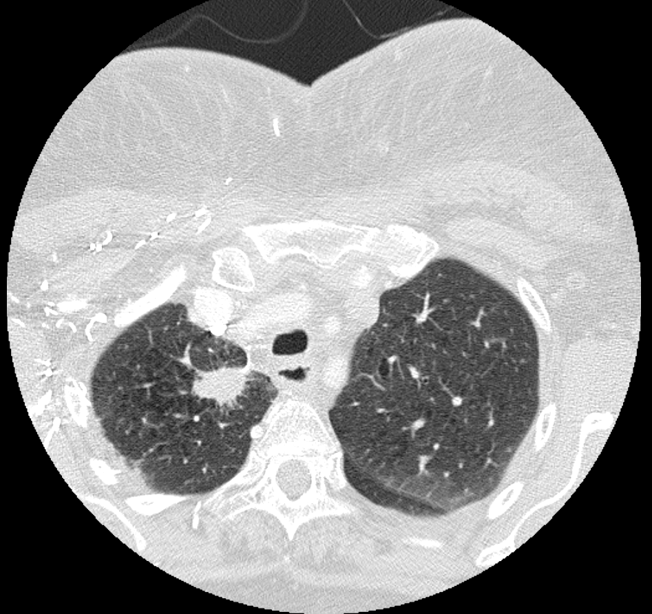
There is a lung mass
There is a large, spiculated lung mass in the right upper lobe.
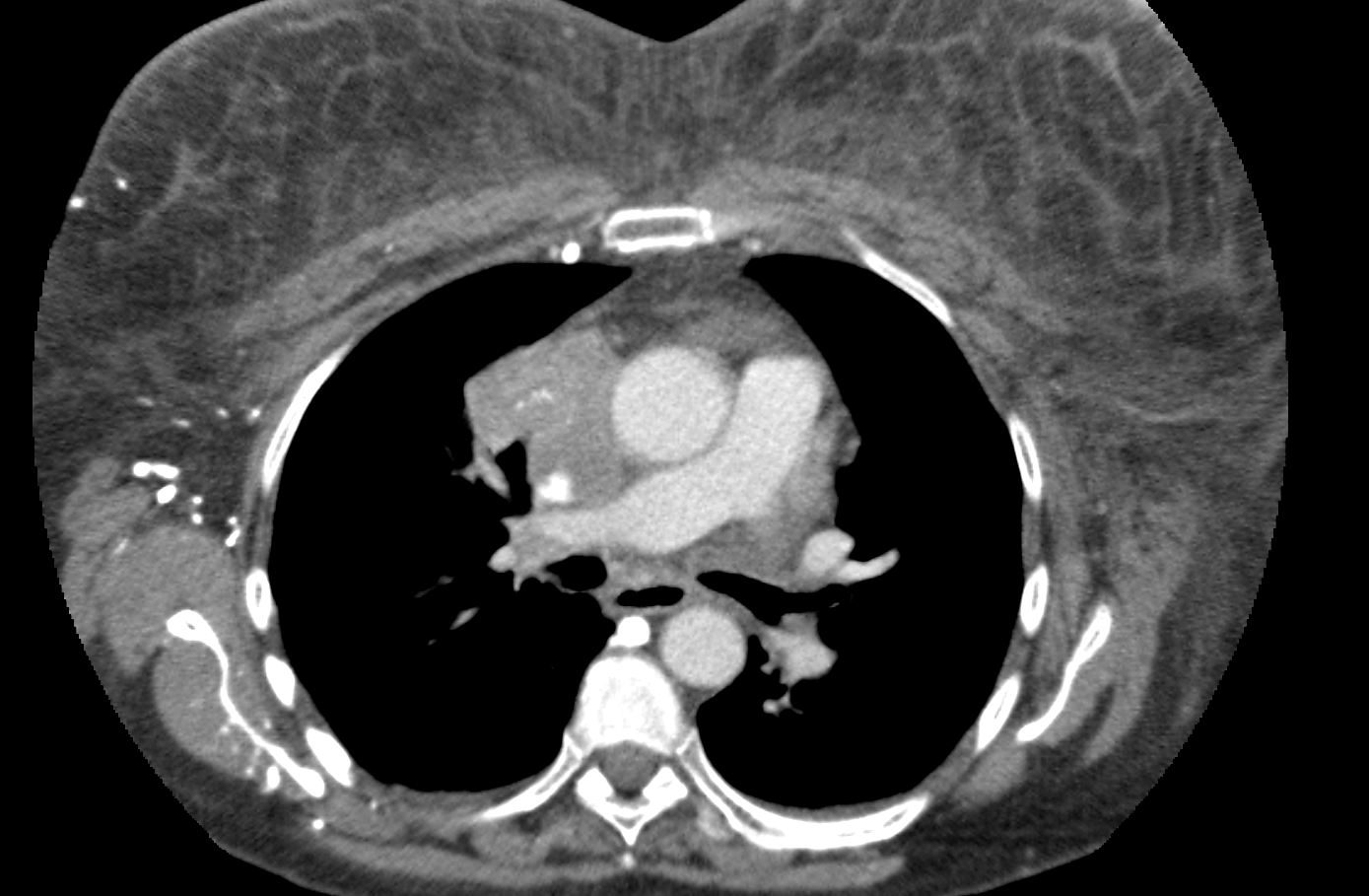
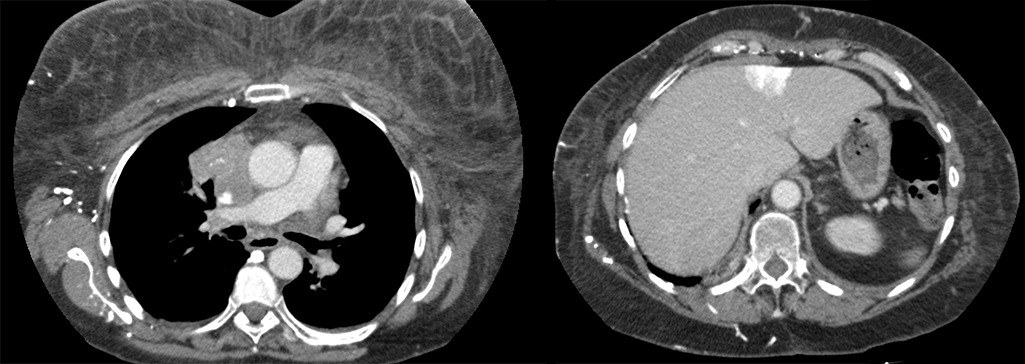
There is evidence of obstruction of the superior vena cava
There is evidence of SVC syndrome considering that there is superior vena cava is compressed by the bulky mediastinal lymphadenopathy, collateral vessels, and the focal hepatic hot spot, or “hot quadrate” sign.
View the full study if you'd like to take a look yourself.
Second Imaging Study
What is the next imaging study you will order?
A staging PET CT would be the next best step. The images are not included for the purposes of this case.
Well done. You were correct
What is your Diagnosis now that you have seen the imaging results?
The clinical and imaging findings are consistent with SVC syndrome secondary to a mass that is likely from metastatic spread of lung cancer to lymph nodes.
Current Acuity
Initially, you selected and we suggested acuity.
Has your concern for this patient changed?
Though the patient’s condition is not immediately life threatening, they will require further workup.
Assessment and Plan
Please provide your assessment and plan for this patient
The patient is a 60-year-old female with significant smoking history presenting with cough, constitutional symptoms, and upper body swelling. Imaging demonstrated right upper lobe masses with a large right mediastinal lymph mass consistent with metastatic disease which encases and occludes the upper SVC, leading to SVC syndrome. Oncology, pulmonology, and vascular surgery should be consulted for further evaluation.
Lessons Learned:
- SVC syndrome can present occur from intrinsic stenosis, thrombosis, or as in this case, extrinsic compression of the SVC.
- SVC syndrome is characterized on CT scan by presence of SVC occlusion, collateral vasculature (including the “hot quadrate” sign), and extensive upper body soft tissue swelling.
That's the end of the module! Once you've reviewed the video(s), you can click here for another case challenge.
Contributors:
Kevin Pierre, MD - Editor
Robbie Slater, MD - Supervising Editor
Bayar Batmunh, MS - Coordinator
Next

