V2) Leg pain after prolonged travel
Review the Learning Outcomes, Hx, PE and Labs, and begin the module with your Provisional Diagnosis. Keep hitting "Next" to move through the module.
Learning Outcomes
- Articulate your relationship with the consulting diagnostic radiologists in the evaluation of a patient with lower extremity pain.
- Review the DDx considerations in a patient with lower extremity pain.
- Identify the spectrum of imaging findings in appropriate modalities for evaluating a patient with lower extremity pain.
History
Physical Exam
Labs
Provisional Diagnosis
Potential Acuity
What is your assessment of the likely acuity for this patient?
First Imaging Study
What is the first imaging study you will order?
Pertinent Imaging Observations
Click on the links below to view images from the study, and assess these key findings as best you can.
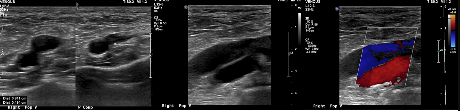
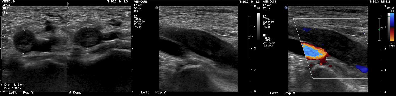
Venous doppler ultrasound of the lower extremities.
There is an occlusive thrombus in the right popliteal vein?
There is an occlusive thrombus in the left popliteal vein?

Watch our video
Second Imaging Study
What is the next imaging study you will order?
What is your Diagnosis now that you have seen the imaging results?
Current Acuity
Initially, you selected and we suggested acuity.
Has your concern for this patient changed?
Assessment and Plan
Please provide your assessment and plan for this patient
Lessons Learned:
- There should be a high clinical suspicion for DVT in patients with components of Virchow’s triad (blood flow stasis, hypercoagulable state, vascular endothelial injury).
- Venous doppler ultrasound, which shows non-compressibility of and lack of flow within the veins, is the gold standard for diagnosing deep venous thromboses.
Socioeconomic Factors: In the US, DVT is most prevalent in African American patients and lowest in Hispanic patients. Insurance status and income level are not associated with the likelihood of admission with a DVT diagnosis. Further research is needed to investigate the cause for these patterns.
That's the end of the module! Once you've reviewed the video(s), you can click here for another case challenge.
Contributors:
Kevin Pierre, MD - Editor
Robbie Slater, MD - Supervising Editor
Bayar Batmunh, MS - Coordinator
