Case Notes
History
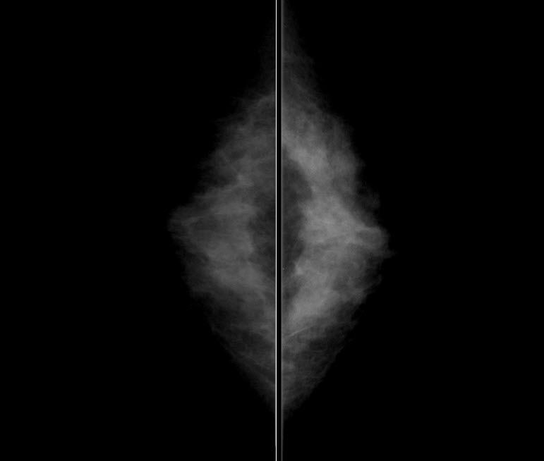
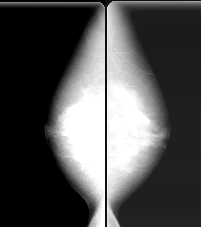
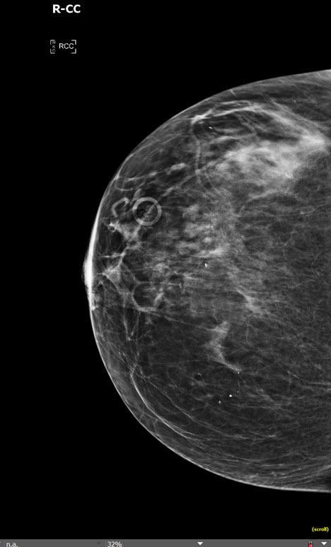
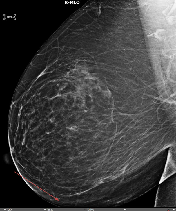
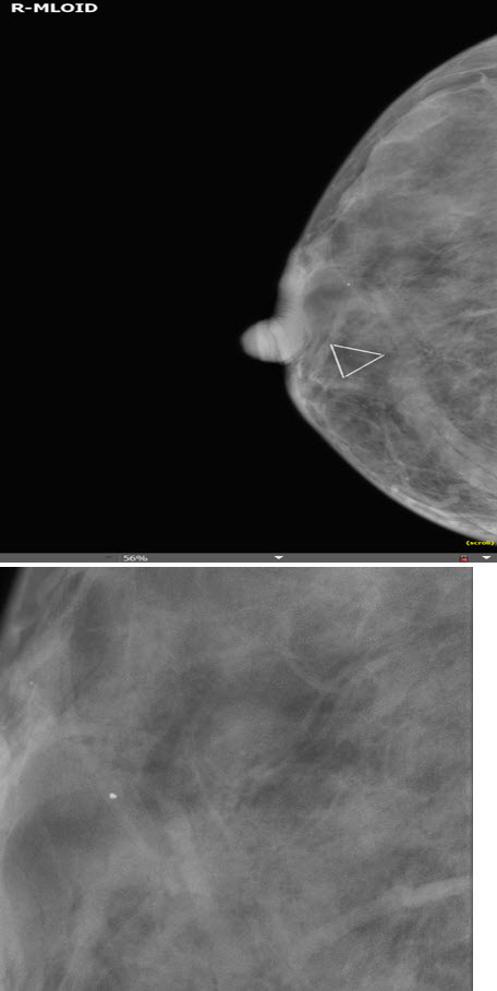
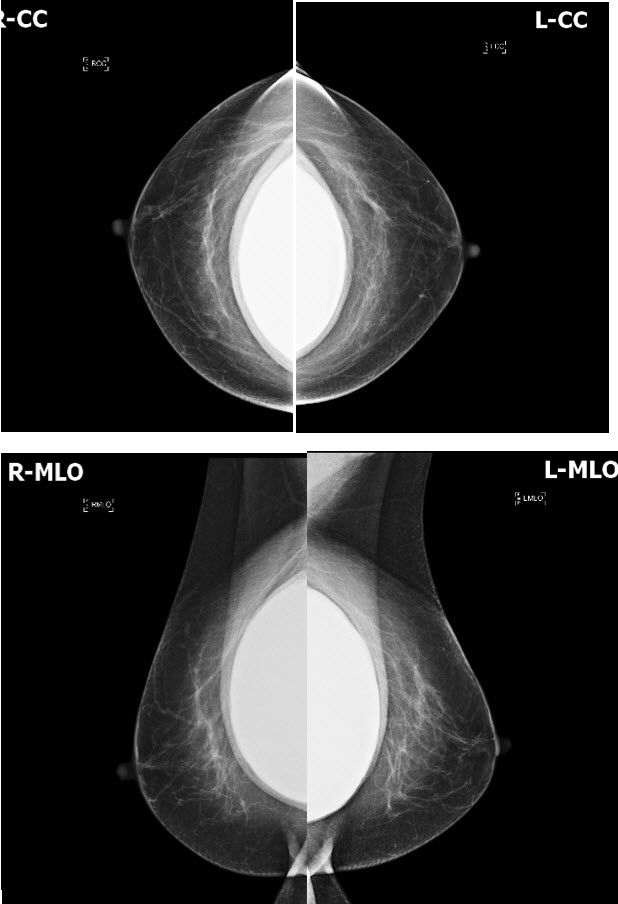
Screening mammogram; the 66 yo patient is asymptomatic.Exam
Prior Study
None availableDicom
Findings
| Technique | Correct Answer | Your Answer |
|---|---|---|
|
|
No | NA |
|
The exam is limited by patient motion. |
No | NA |
|
The exam is limited by exclusion of tissue. |
No | NA |
|
The exam is limited by artifact. |
No | NA |
| Breast Density | Correct Answer | Your Answer |
|---|---|---|
|
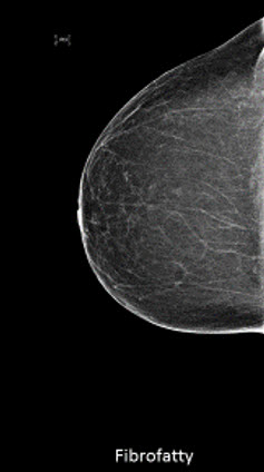
There is a fibrofatty glandular pattern. |
No | NA |
|
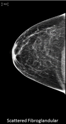
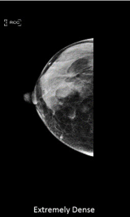
There is a scattered fibroglandular pattern. |
Yes | NA |
|
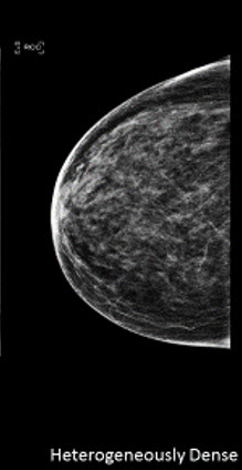
There is a heterogeneously dense fibroglandular pattern. |
No | NA |
|
There is an extremely dense fibroglandular pattern. |
No | NA |
| Mass | Correct Answer | Your Answer |
|---|---|---|
|
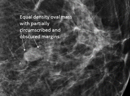
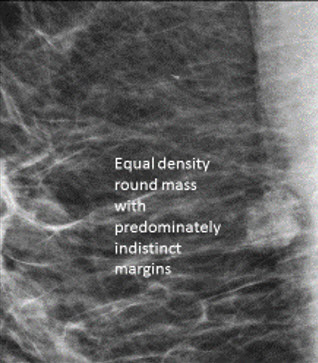
There is a mass (masses) present. |
No | NA |
|
The mass is right/left/bilateral. |
N/A | NA |
|
The mass is new. |
N/A | NA |
|
The mass is stable. |
N/A | NA |
|
The mass is low density relative to the breast tissue |
N/A | NA |
|
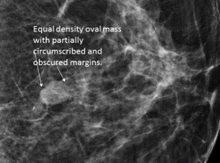
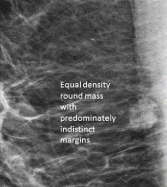
The mass is equal density relative to the breast tissue |
N/A | NA |
|
The mass is high density relative to the breast tissue. |
N/A | NA |
|
The mass contains fat. |
N/A | NA |
|
|
N/A | NA |
|
There are associated calcifications. |
N/A | NA |
|
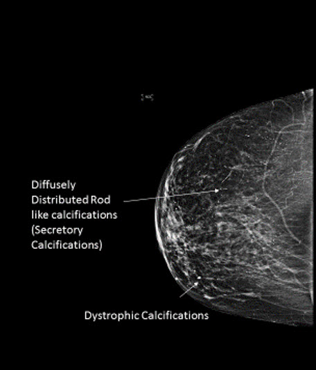
The calcifications are dystrophic. |
N/A | NA |
|
There are benign intramammary lymph nodes. |
No | NA |
| Margins | Correct Answer | Your Answer |
|---|---|---|
|
The margins of the mass are circumscribed. |
N/A | NA |
|
The margins of the mass are obscured. |
N/A | NA |
|
The margins of the mass are indistinct. |
N/A | NA |
|
The margins of the mass are spiculated. |
N/A | NA |
| Calcifications | Correct Answer | Your Answer |
|---|---|---|
|
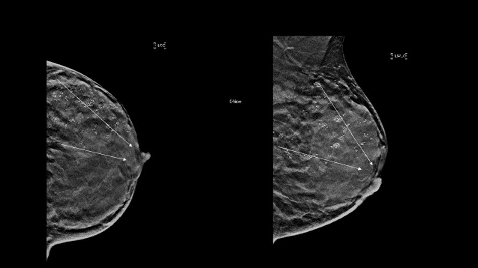
There are calcifications. |
Yes | NA |
|
The calcifications are right/left/bilateral |
Yes | NA |
|
The calcifications are new. |
No | NA |
|
The calcifications are stable. |
N/A | NA |
|
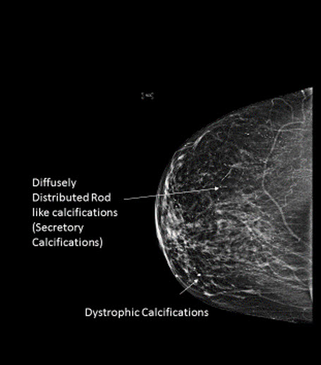
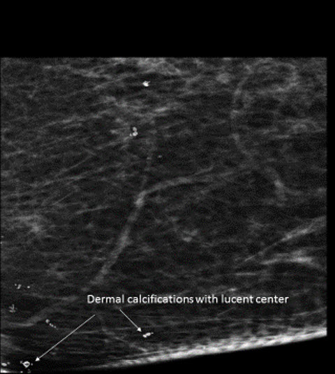
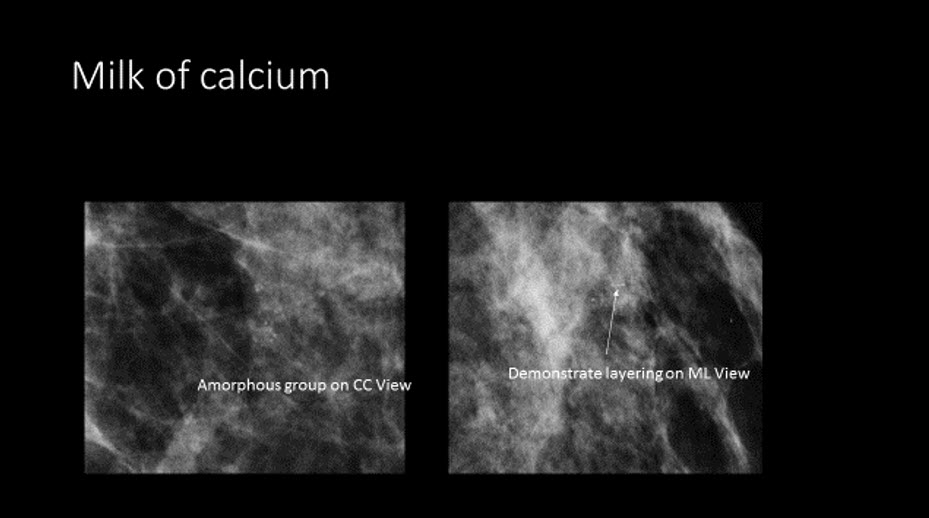
The calcifications are benign (vascular, coarse dystrophic, secretory, skin, milk of calcium). |
Yes | NA |
|
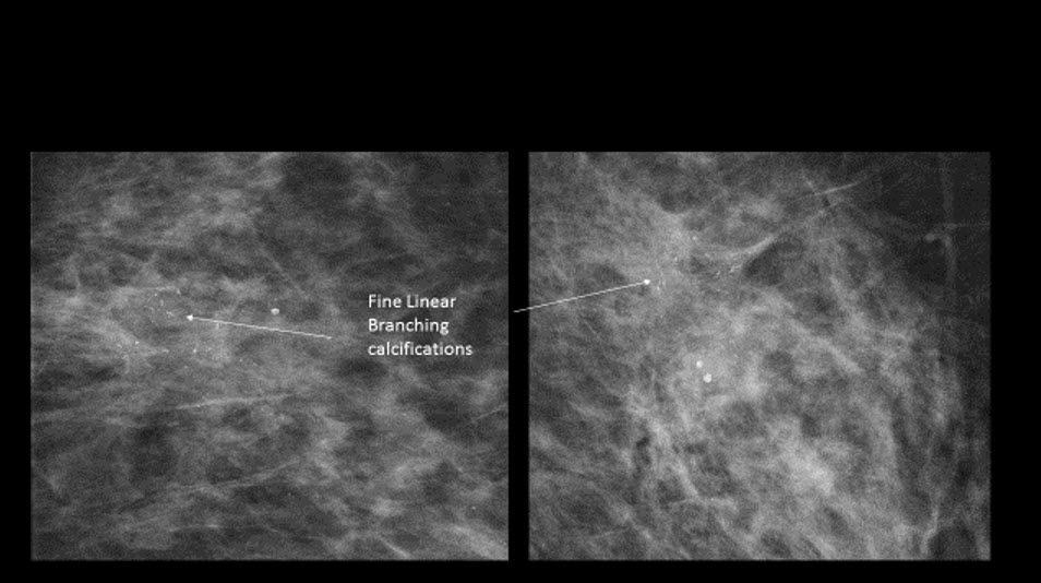
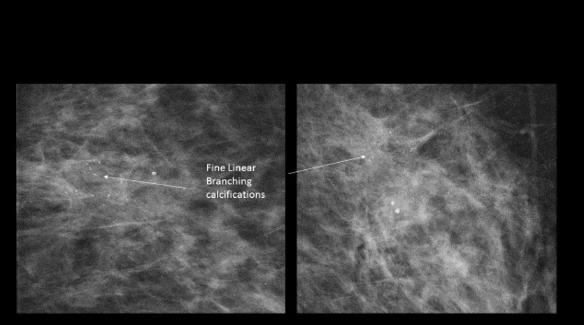
The calcifications are suspicious (amorphous, coarse heterogeneous, fine pleomorphic, fine linear, fine linear and branching). |
No | NA |
|
The calcifications are diffusely distributed. |
Yes | NA |
|
The calcifications are regionally distributed. |
No | NA |
|
The calcifications are grouped. |
No | NA |
|
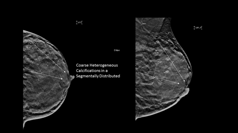
The calcifications are segmentally distributed. |
No | NA |
|
The calcifications are linearly distributed. |
No | NA |
| Asymmetry | Correct Answer | Your Answer |
|---|---|---|
|
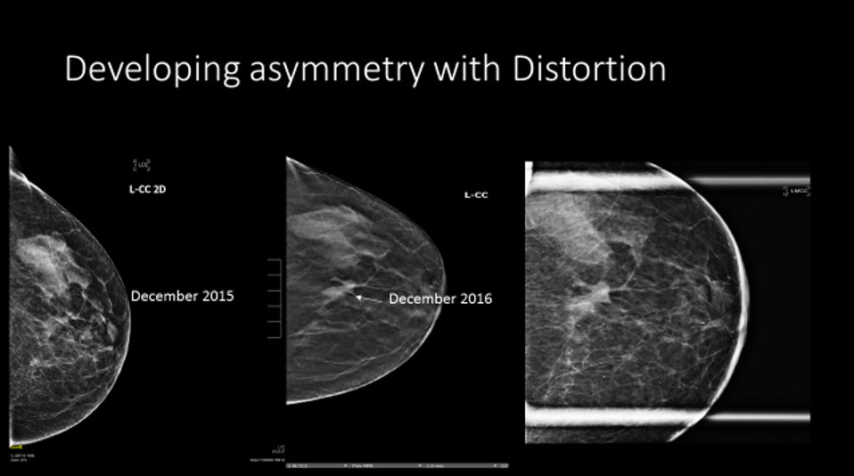
There is an asymmetry (focal, regional, global). |
No | NA |
|
The asymmetry is right/left/bilateral. |
N/A | NA |
|
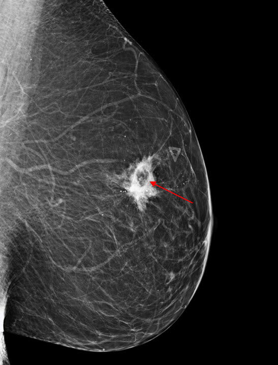
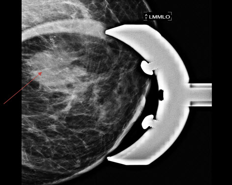
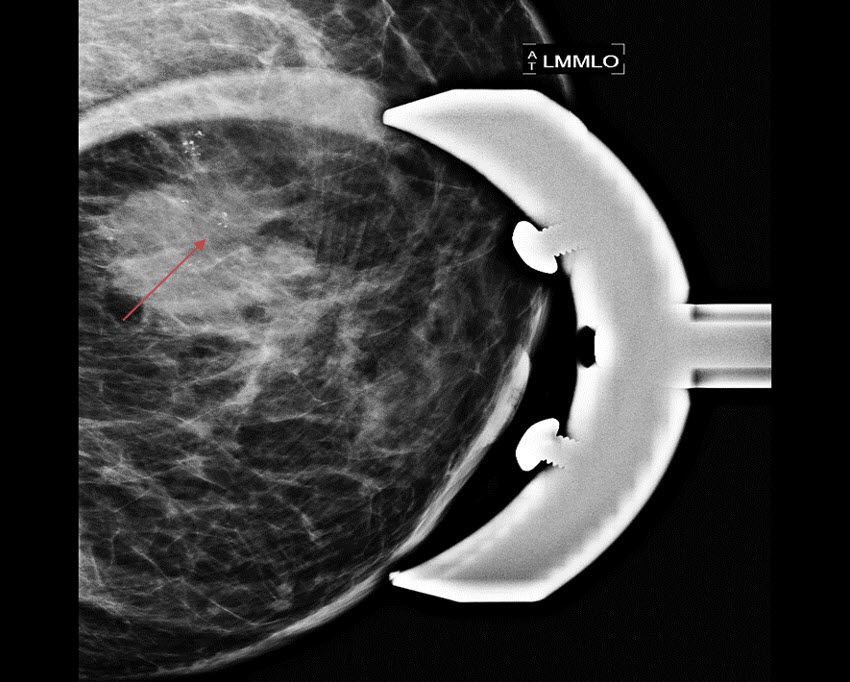
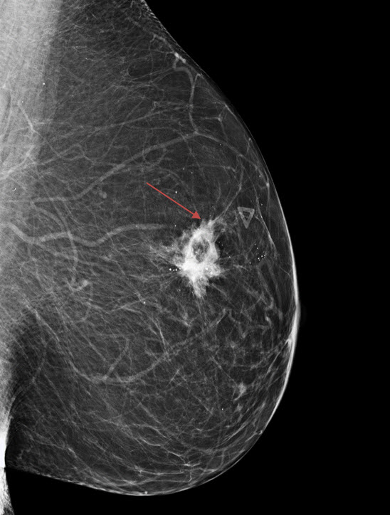
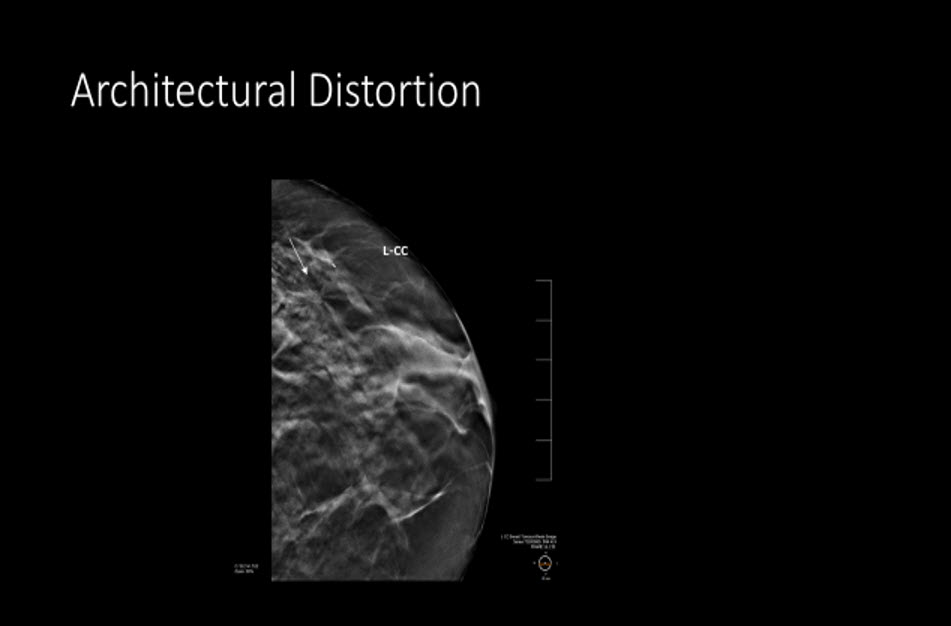
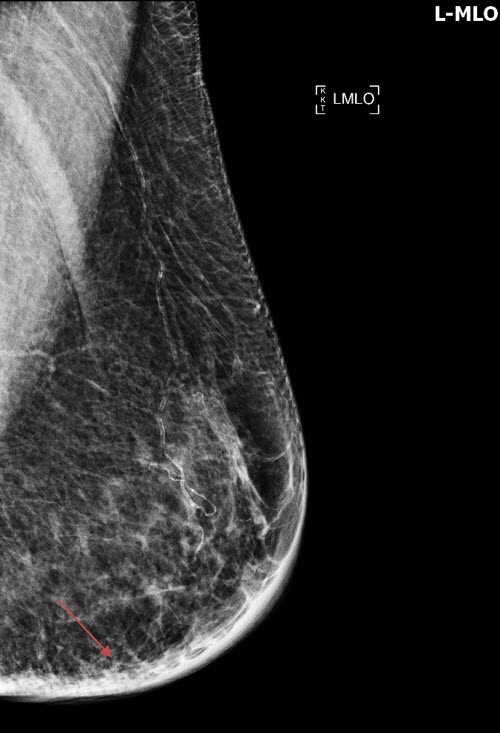
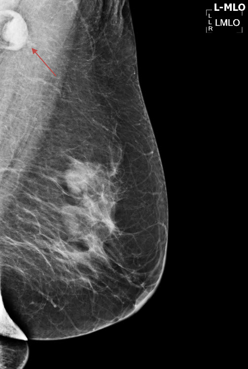
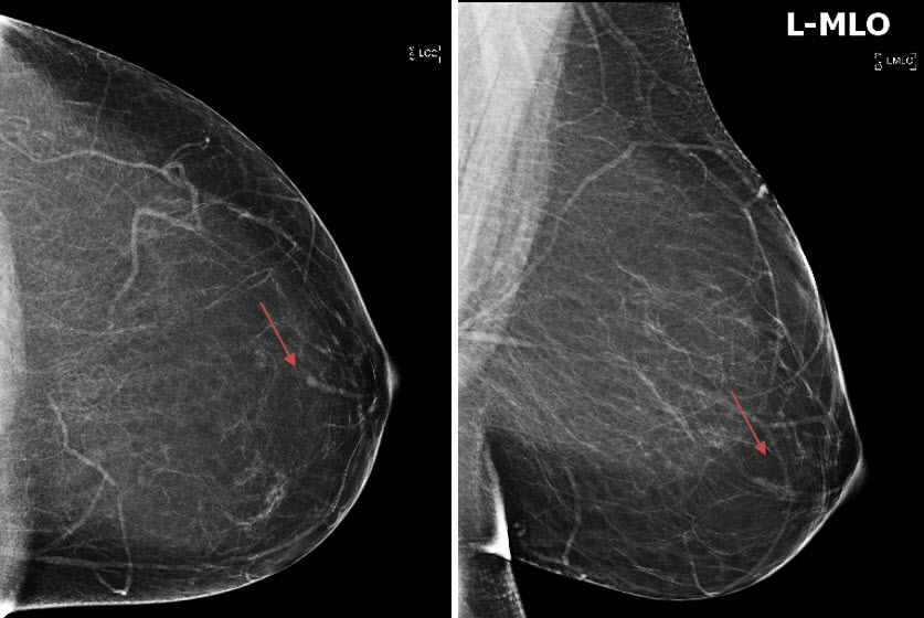
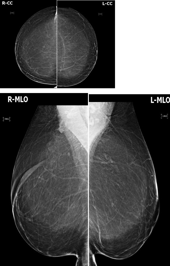
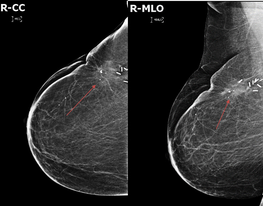
There is architectural distortion. |
Yes | NA |
|
The architectural distortion is right/left/bilateral. |
Yes | NA |
| Associated Features | Correct Answer | Your Answer |
|---|---|---|
|
There is nipple retraction. |
No | NA |
|
The nipple retraction is right/left/bilateral. |
N/A | NA |
|
There is skin thickening. |
No | NA |
|
The skin thickening is right/left/bilateral. |
N/A | NA |
| Axilla | Correct Answer | Your Answer |
|---|---|---|
|
There are morphologically abnormal lymph nodes. |
No | NA |
|
The morphologically abnormal lymph nodes are right/left/bilateral. |
N/A | NA |
| Subareoalar Region | Correct Answer | Your Answer |
|---|---|---|
|
There are abnormal ducts in the subareolar region. |
No | NA |
|
The abnormal ducts in the subareolar region are right/left/bilateral. |
N/A | NA |
| Prior Surgery | Correct Answer | Your Answer |
|---|---|---|
|
There are implants. |
No | NA |
|
The implants are subglandular/subpectoral. |
N/A | NA |
|
The implants are saline/silicone. |
N/A | NA |
|
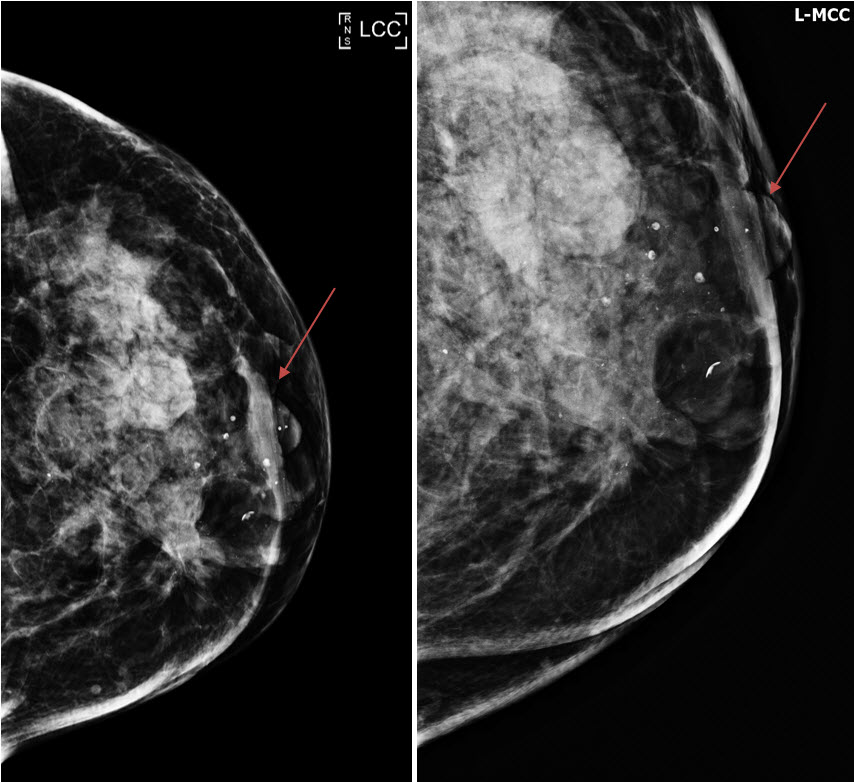
There are changes related to prior surgery breast reduction/lumpectomy/surgical excision. |
Yes | NA |
|
The changes related to prior surgery are right/left/bilateral. |
Left | NA |
Impression
Expert Answer
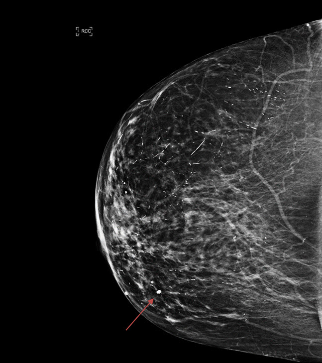
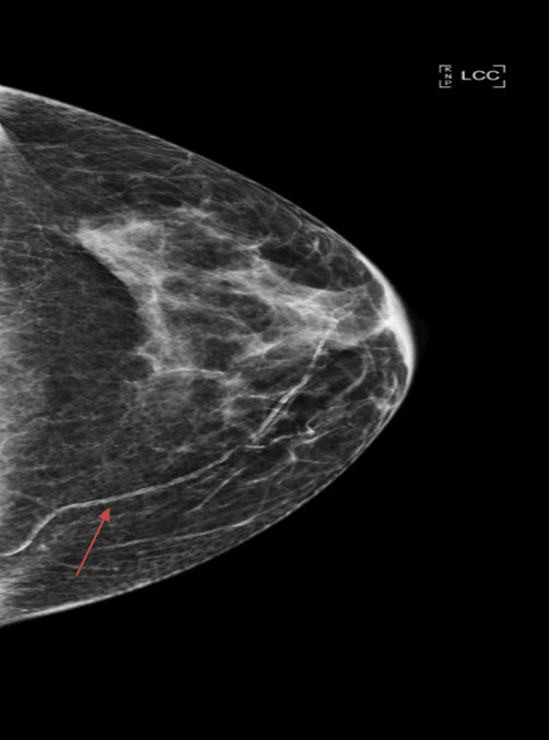
There is architectural distortion in the upper outer right breast. BI-RADS Category 0: Mammography: Incomplete – Need Additional Imaging Evaluation and/or Prior Mammograms for Comparison
Your Answer
Recommendations & Acuity
Recommendations
Expert Answer
Spot compression views and targeted ultrasound of the upper outer right breast are recommended.
Your Answer
Acuity
Expert Answer
Emergent (Action Necessary now)
