Retake
N34) Progressive headaches and unsteadiness in a 25-year-old male
Review the Learning Outcomes, Hx, PE and Labs, and begin the module with your Provisional Diagnosis. Keep hitting "Next" to move through the module.
Learning Outcomes
- Articulate your relationship with the consulting diagnostic radiologists in the evaluation of a patient with headache.
- Review the DDx considerations in a patient with headache.
- Identify the spectrum of imaging findings in appropriate modalities for evaluating patients with headache.
History
A 25-year-old male presents to his primary care provider with a 6-month history of progressive headaches that worsen when straining. He also reports unsteadiness when walking. He has also had episodes of difficulty swallowing and numbness tingling in his hands and arms. He denies any past medical history other than a left mastoidectomy.
Physical Exam
BP: 115/73, HR 81, RR 17, Temp 37.6 C (98 F), O2 saturation 99%.
Neurologic Exam: Downbeat nystagmus. 3+ deep tendon reflexes in bilateral upper and lower extremities. Positive Romberg sign, bilateral sensorineural hearing loss, and 4+/5 strength and decreased pain and temperature sensations in bilateral upper extremities. There is dysmetria and dysdiadochokinesia.
Labs
None
Provisional Diagnosis
Select the Dx you believe is most appropriate
A Chiari malformation is the most likely diagnosis in this patient with progressive headaches that worsen with maneuvers that increase ICP, difficulty swallowing, upper extremity bilateral sensory symptoms and weakness, and cerebellar symptoms (dysmetria and dysdiadochokinesia).
Well done. You were correct
Potential Acuity
What is your assessment of the likely acuity for this patient?
Well done. You were correct
The patient requires routine, but expedited workup.
First Imaging Study
What is the first imaging study you will order?
An MRI of the brain is an appropriate imaging modality as it can evaluate the position of the cerebellar tonsils.
Well done. You were correct
Pertinent Imaging Observations
Click on the links below to view images from the study, and assess these key findings as best you can.
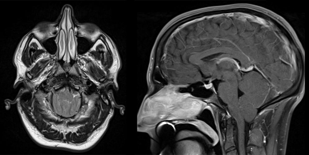
MRI brain
The cerebellum is normal.
There is herniation of the cerebellar tonsils below the foramen magnum.
View the full study if you'd like to take a look yourself.
Second Imaging Study
What is the next imaging study you will order?
No further imaging is necessary as the diagnosis is made using the MRI.
Well done. You were correct
What is your Diagnosis now that you have seen the imaging results?
A Type I Chiari malformation is the diagnosis. Type II Chiari malformation would present with herniation of the vermis in addition to the cerebellar tonsils and is associated with myelomeningoceles.
Current Acuity
Initially, you selected and we suggested acuity.
Has your concern for this patient changed?
The patient requires routine, but expedited workup.
Assessment and Plan
Please provide your assessment and plan for this patient
A 25-year-old male presents with symptoms, physical exam, and imaging findings consistent with a Chiari I malformation. He should be referred to a neurosurgeon for consideration for a posterior fossa decompression.
Lessons Learned:
- Chiari Malformation Type I is characterized by a herniation of the cerebellar tonsils through the foramen magnum, which can lead to compression of the brainstem.
- Clinical presentations often involve bulbar and ocular symptoms.
- Chiari malformations are also associated with the presence of a syrinx.
- Cross-sectional imaging can be used for diagnosis. The key finding is tonsillar ectopia 3mm below McRae’s line, which is drawn between the opisthion and the basion.
That's the end of the module! Once you've reviewed the video(s), you can click here for another case challenge.
Next

