Retake
N19) Fever, facial pain, and jaw swelling
Review the Learning Outcomes, Hx, PE and Labs, and begin the module with your Provisional Diagnosis. Keep hitting "Next" to move through the module.
Learning Outcomes
- Articulate your relationship with the consulting diagnostic radiologists in the evaluation of a patient with facial swelling and fever.
- Review the DDx considerations in a patient with facial swelling and fever.
- Identiy the spectrum of imaging findings in appropriate modalities for evaluating patients with facial swelling and fever.
History
A 50-year old male presents due to a progressively worsening fever, trismus and facial pain. He reports having noticed swelling on the left side of his face near the jaw about a week ago, which has been increasingly painful. He also describes difficulty opening his mouth and swallowing.
Physical Exam
BP: 142/89, HR: 82 bpm, RR: 19, Temp: 102F, O2 saturation: 97%.
HEENT: Poor oral hygiene. Expression of purulent exudate from Stensen’s duct with palpation of the parotid gland. Left-sided facial swelling with tenderness on palpation of the parotid gland. Left-sided tender cervical lymphadenopathy. The patient has difficulty opening his mouth.
Labs
White blood cells (WBC): 14,000/uL (Reference range: 4,500-11,000/uL),
Neutrophils: 82% (Reference range: 40-70%)
Provisional Diagnosis
Select the Dx you believe is most appropriate
This patient most likely has a parotid gland infection considering the left sided parotid tenderness, purulent exudate from Stensen’s duct, fever, and leukocytosis with a neutrophilic predominance.
Well done. You were correct
Potential Acuity
What is your assessment of the likely acuity for this patient?
Well done. You were correct
This patient requires urgent work up for his condition.
First Imaging Study
What is the first imaging study you will order?
An ultrasound is an appropriate initial imaging modality; however, the quality of a salivary gland ultrasound is highly dependent upon operator technique, and in facilities with less experience and expertise in salivary gland ultrasound, a CT with contrast is an alternative first-line imaging modality. In this case, a CT will be performed. A CT with contrast is most sensitive in determining the depth and extent of an abscess and in detecting involvement of surrounding structures.
Well done. You were correct
Pertinent Imaging Observations
Click on the links below to view images from the study, and assess these key findings as best you can.
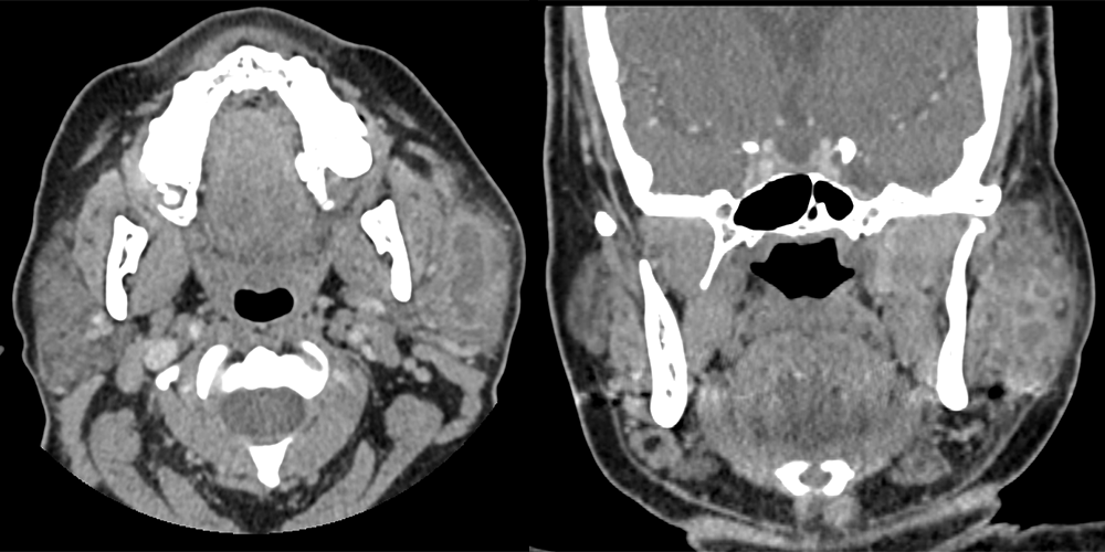
Maxillofacial CT scan
The left parotid gland is inflamed.
There is extensive inflammatory disease involving the left parotid gland, or sialadenitis, as evidenced by contrast enhancement with adjacent fat stranding.
The left parotid gland is otherwise normal
There is a loculated abscess in the left parotid gland, as evidenced by its peripheral enhancement and hypodense center.
View the full study if you'd like to take a look yourself.
Second Imaging Study
What is the next imaging study you will order?
No further imaging is required as the diagnosis is made with the CT scan.
Well done. You were correct
What is your Diagnosis now that you have seen the imaging results?
The patient likely has obstruction of the left parotid duct as evidenced by the dilated left parotid duct. This obstruction likely allowed for an infection to occur, resulting in parotid gland abscess and sialadenitis.
Current Acuity
Initially, you selected and we suggested acuity.
Has your concern for this patient changed?
The patient requires urgent management.
Assessment and Plan
Please provide your assessment and plan for this patient
This patient is a 50-year-old male presenting with a parotid gland abscess secondary to obstruction with associated sialadenitis, reactive adenopathy, and surrounding cellulitis. He will require hospital admission, a consultation with an ENT or Radiology for potential abscess drainage—with aspirations sent for culture analyses. Hydration, administration of analgesics for pain control, and delivery of intravenous broad-spectrum antibiotics until the culture results are obtained are required.
Lessons Learned:
- Acute sialadenitis is characterized by sudden enlargement and pain of the affected gland and is usually due to an obstructive, infectious, or inflammatory etiology.
- Signs and symptoms of a bacterial infection in the setting of obstruction include increasing gland size and pain, and development of purulent saliva and fever.
- The parotid gland drains into the mouth via the Stensen’s duct near the second upper molar.
- CT with contrast is an alternative first-line imaging to ultrasound.
- Initial treatment should be in an inpatient setting, since suppurative parotitis may spread to deep fascial spaces of the head and neck.
Socioeconomic Factors: Acute bacterial parotitis is more commonly found in elderly patients, likely because they often take medications with an atropine effect causing reduced salivary flow and predisposing them to a retrograde infection.
That's the end of the module! Once you've reviewed the video(s), you can click here for another case challenge.
Next

