N15) New onset seizure in a patient with headaches
Review the Learning Outcomes, Hx, PE and Labs, and begin the module with your Provisional Diagnosis. Keep hitting "Next" to move through the module.
Learning Outcomes
- Articulate your relationship with the consulting diagnostic radiologists in the evaluation of a patient with seizures.
- Review the DDx considerations in a patient with seizures.
- Identify the spectrum of imaging findings in appropriate modalities for evaluating patients with seizures.
History
Physical Exam
Labs
Provisional Diagnosis
Potential Acuity
What is your assessment of the likely acuity for this patient?
First Imaging Study
What is the first imaging study you will order?
Pertinent Imaging Observations
Click on the links below to view images from the study, and assess these key findings as best you can.
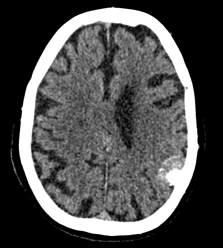
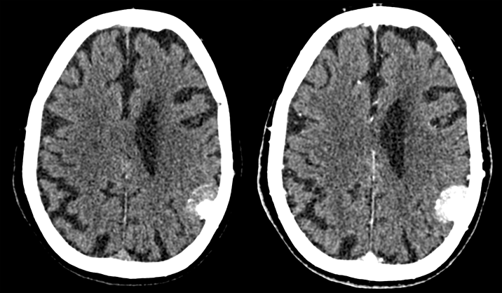
CT head with and without contrast
There is a space occupying lesion.
The space occupying lesion is in which lobe?
There are intra-lesional calcifications.
The lesion is enhancing.

Watch our video
Second Imaging Study
What is the next imaging study you will order?
Pertinent Imaging Observations
Click on the links below to view images from the study, and assess these key findings as best you can.
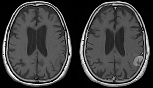
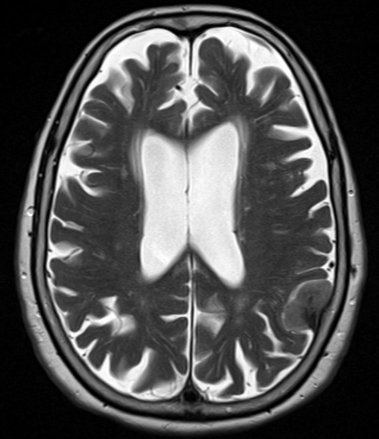
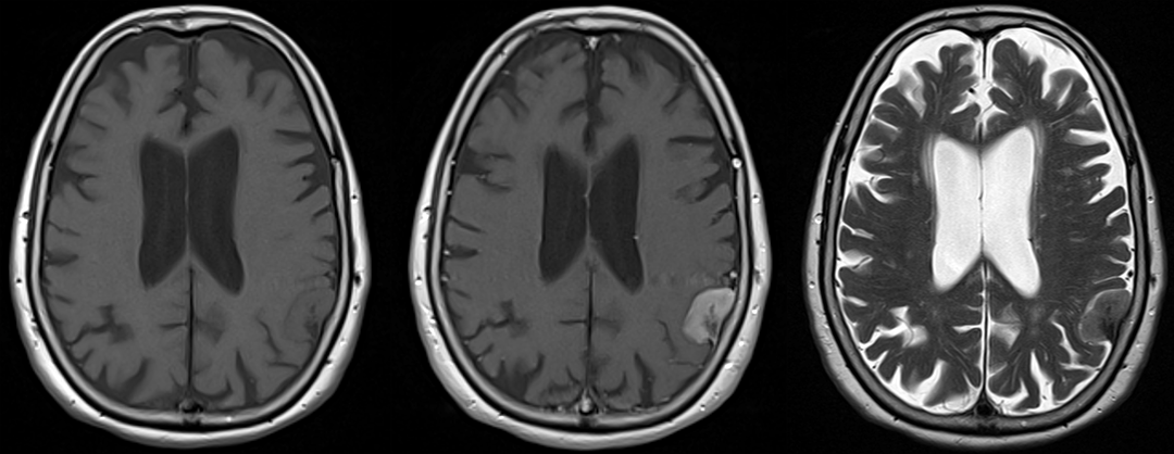
MRI
The lesion is homogeneously enhancing.
The lesion is
Which of the following describes the lesion characteristics?
Watch our video
What is your Diagnosis now that you have seen the imaging results?
Current Acuity
Initially, you selected and we suggested acuity.
Has your concern for this patient changed?
Assessment and Plan
Please provide your assessment and plan for this patient
Lessons Learned:
- Meningiomas are extra-axial masses that arise from meningothelial cells. Risk factors include older age, female sex, previous exposure to ionizing radiation, and genetic predisposition (NF2 and schwannomatosis).
- Symptoms from mass effect may include headaches, seizures and focal neurological deficits.
- CT and MRI classically reveals an enhancing, calcified, extra-axial mass.
- Imaging signs associated with meningioma include CSF cleft sign, a dural tail, and adjacent hyperostosis (especially on the skull base).
- Pathology, which would show ‘fried egg’ cells, psammoma bodies, and Rosenthal fibers confirm the diagnosis.
- Resection only is considered curative for WHO grade I tumors.
Socioeconomic Factors:
- Patients with lower socioeconomic status have lower rates of gross total resection and survival.
That's the end of the module! Once you've reviewed the video(s), you can click here for another case challenge.
Contributors:
Abeer Dagra, MS3 - Content Contributor
Kevin Pierre, MD - Editor
Robbie Slater, MD - Supervising Editor
Bayar Batmunh, MS - Coordinator
