Retake
N13) Sudden-onset weakness and slurred speech
Review the Learning Outcomes, Hx, PE and Labs, and begin the module with your Provisional Diagnosis. Keep hitting "Next" to move through the module.
Learning Outcomes
- Articulate your relationship with the consulting diagnostic radiologists in the evaluation of a patient with focal neurologic deficits.
- Review the DDx considerations in patients presenting with focal neurologic deficits.
- Identify the spectrum of imaging findings in appropriate modalities for evaluating patients with focal neurologic deficits.
History
A 65-year-old male presents to the emergency department due to sudden onset left-sided weakness and slurred speech that began about an hour ago. He denies any recent trauma. He has a history of hypertension, type 2 diabetes mellitus, and hyperlipidemia, but has not been compliant with his medications.
Physical Exam
BP: 190/110, HR 100, RR 22, Temp 99.3 F, O2 saturation 96%.
Neurological examination: Alert and oriented to person, place, and time; cranial nerves II-XII intact; left-sided facial droop; motor strength 2/5 in left upper and lower extremities, 5/5 on the right side; sensation intact bilaterally.
Labs
None
Provisional Diagnosis
Select the Dx you believe is most appropriate
Given the patient's uncontrolled hypertension, sudden onset of focal neurologic deficits, slurred speech, and left-sided facial droop, the most appropriate provisional diagnosis is a stroke. The location is likely in the right basal ganglia, considering the contralateral hemiparesis and facial droop. A stroke in the right middle cerebral artery (MCA) or anterior cerebral artery (ACA) may present with contralateral weakness, with the lower extremity relatively spared in the ACA and the upper extremity relatively spared in the MCA. A posterior cerebral artery (PCA) stroke might result in visual field deficits. A cerebellar stroke would typically present with ipsilateral ataxia and nystagmus.
Well done. You were correct
Potential Acuity
What is your assessment of the likely acuity for this patient?
Well done. You were correct
A stroke necessitates immediate evaluation and management, as delays in care can lead to irreversible brain damage and poorer outcomes, emphasizing the adage, "time is brain."
First Imaging Study
What is the first imaging study you will order?
A head CT without contrast is a rapidly obtainable test that can effectively rule out an intracranial hemorrhage.
Well done. You were correct
Pertinent Imaging Observations
Click on the links below to view images from the study, and assess these key findings as best you can.
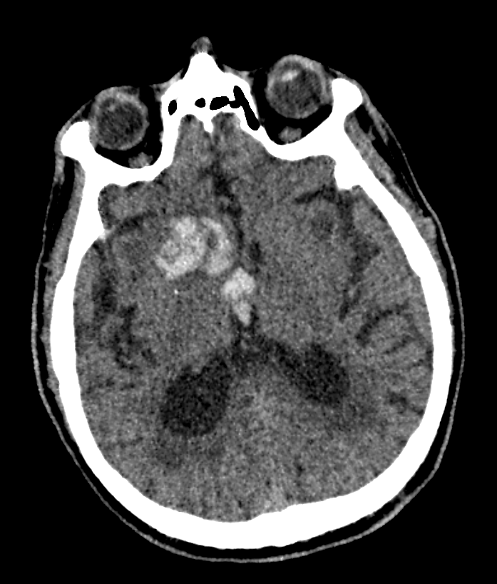
Head CT without contrast
What type of intracranial hemorrhage is present?
The hemorrhage is observed within the brain parenchyma, consistent with an IPH, and not in the cisterns (SAH) or associated with the dura (EDH vs SDH).
Where is the intracranial hemorrhage located?
The hemorrhage can be seen in the deep brain structures of the basal ganglia, which is the classic location for a hypertensive stroke.
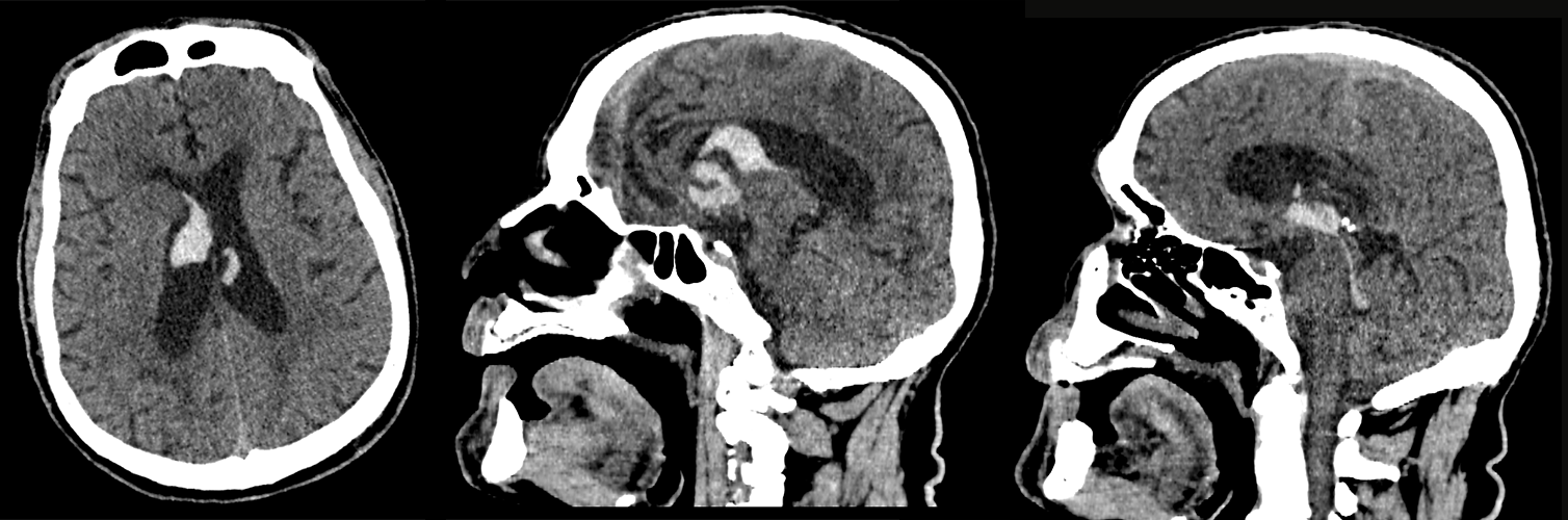
Is there intraventricular extension?
The blood products are seen in the ventricular space as well, indicating intraventricular extension.
How far does it extend?
The blood extends into the 4th ventricle.
View the full study if you'd like to take a look yourself.
Pertinent Imaging Observations
Click on the links below to view images from the study, and assess these key findings as best you can.
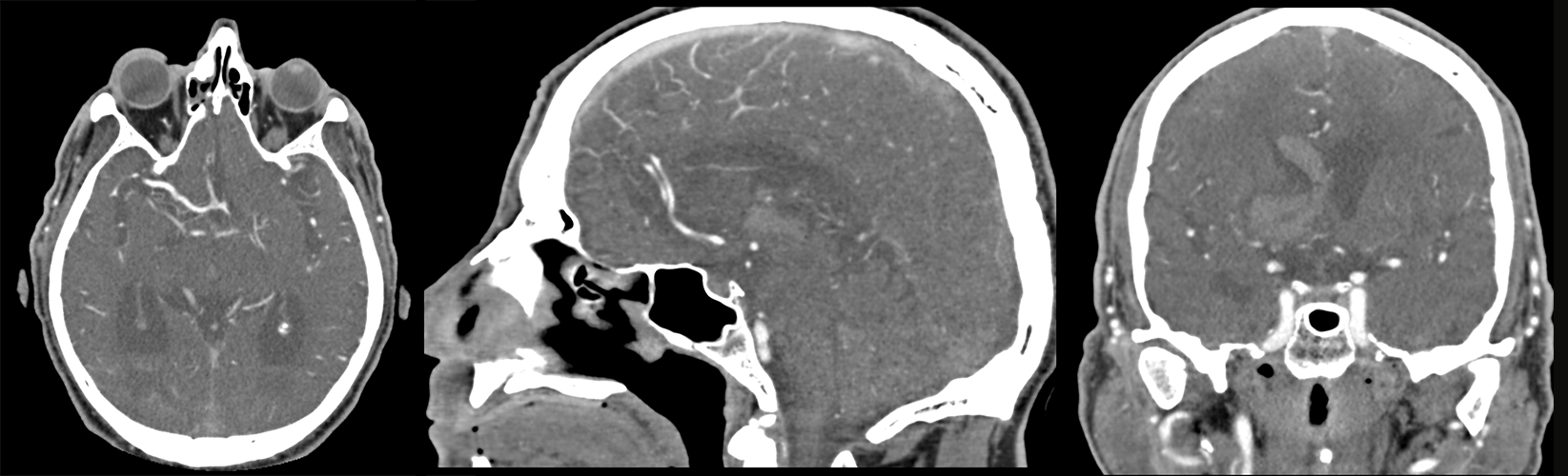
CTA of the head and neck with IV contrast
Is there evidence of an aneurysm?
There is no aneurysm on the imaging.
View the full study if you'd like to like a look yourself
What is your Diagnosis now that you have seen the imaging results?
Considering the patient's history of uncontrolled hypertension and the absence of an arteriovenous malformation or aneurysm on the CT scan, their intracranial hemorrhage is likely attributable to hypertension.
Current Acuity
Initially, you selected and we suggested acuity.
Has your concern for this patient changed?
The acuity may be downgraded from emergent to urgent in the case of a hemorrhagic stroke because the management strategies are different compared to an ischemic stroke. Hemorrhagic strokes do not have the same time-sensitive thrombolytic treatment options as ischemic strokes, and the focus shifts to blood pressure control, supportive care, and addressing the underlying cause to prevent further bleeding and complications.
Assessment and Plan
Please provide your assessment and plan for this patient
The 65-year-old male patient with a history of uncontrolled hypertension presents with a right basal ganglia intraparenchymal hemorrhage, likely secondary to hypertension. He requires ICU monitoring, blood pressure management with IV antihypertensives, and neurosurgical consultation. A neurology consult will be obtained for further evaluation, and physical and occupational therapy consultations will be requested to initiate appropriate rehabilitation interventions. Labs will be monitored closely, and the patient and his family will be counseled on medication adherence and lifestyle modifications to reduce the risk of future strokes.
Lessons Learned:
- Intraparenchymal hemorrhage, also known as hemorrhagic stroke, involves bleeding within the brain tissue due to causes such as trauma, stroke, aneurysms, or hypertension. Identifying the etiology is crucial for management.
- A non-contrast CT can confirm the diagnosis and evaluate the location and extent of the bleeding. Subsequently, a minimally invasive imaging technique, like a CTA or MRA, can be employed to identify a vascular origin for the hemorrhagic stroke with higher sensitivity.
- Management typically includes blood pressure regulation, monitoring and managing intracranial pressure, providing supportive care (e.g., oxygen and fluids), and addressing underlying medical conditions. In some instances, neurosurgical intervention may be needed to remove the blood clot or repair the responsible blood vessel.
- Prognosis for intracranial hemorrhage depends on various factors, such as the cause and severity, location of the bleed, and the patient's overall health status.
That's the end of the module! Once you've reviewed the video(s), you can click here for another case challenge.
Next

