Retake
N11) Acute-onset weakness and slurred speech
Review the Learning Outcomes, Hx, PE and Labs, and begin the module with your Provisional Diagnosis. Keep hitting "Next" to move through the module.
Learning Outcomes
- Articulate your relationship with the consulting diagnostic radiologists in the evaluation of a patient with weakness.
- Review the DDx considerations in a patient with weakness.
- Identify the spectrum of imaging findings in appropriate modalities for evaluating patients with weakness.
History
An 85-year-old male patient with a medical history of hypertension, hyperlipidemia, type 2 diabetes mellitus, atrial fibrillation, and 60 pack-years of smoking presents with acute onset right-sided weakness and slurred speech, which started two hours ago. The patient often does not take their medication. He denies any headaches.
Physical Exam
BP: 154/85, HR 98 bpm, RR 20, Temp 98.6, O2 saturation 98%. Cardiovascular: Irregularly irregular rhythm. No murmurs, rubs, or gallops. Right lower extremity strength 4/5, right upper extremity strength 2/5. Decreased sensation to touch on the right side. Right-sided facial droop. +3 right brachioradialis deep tendon reflex. NIHSS score of 7.
Labs
None
Provisional Diagnosis
Select the Dx you believe is most appropriate
Considering the patient's significant risk factors (hypertension, hyperlipidemia, type 2 diabetes mellitus, and atrial fibrillation), the presentation is most likely a cerebrovascular accident. The left MCA territory is the probable location, given the right-sided neurological deficits, which are more pronounced in the upper extremities. The MCA supplies the lateral part of the cerebral hemisphere, including the primary motor and sensory cortex areas responsible for the face and upper extremities. An ACA stroke would typically have more severe manifestations in the lower extremities.
Well done. You were correct
Potential Acuity
What is your assessment of the likely acuity for this patient?
Well done. You were correct
A stroke necessitates immediate evaluation and management, as delays in care can lead to irreversible brain damage and poorer outcomes, emphasizing the adage, "time is brain."
First Imaging Study
What is the first imaging study you will order?
A head CT without contrast is a rapidly obtainable test that can effectively rule out an intracranial hemorrhage.
Well done. You were correct
Pertinent Imaging Observations
Click on the links below to view images from the study, and assess these key findings as best you can.
Head CT without contrast
Is there evidence of intracranial hemorrhage?
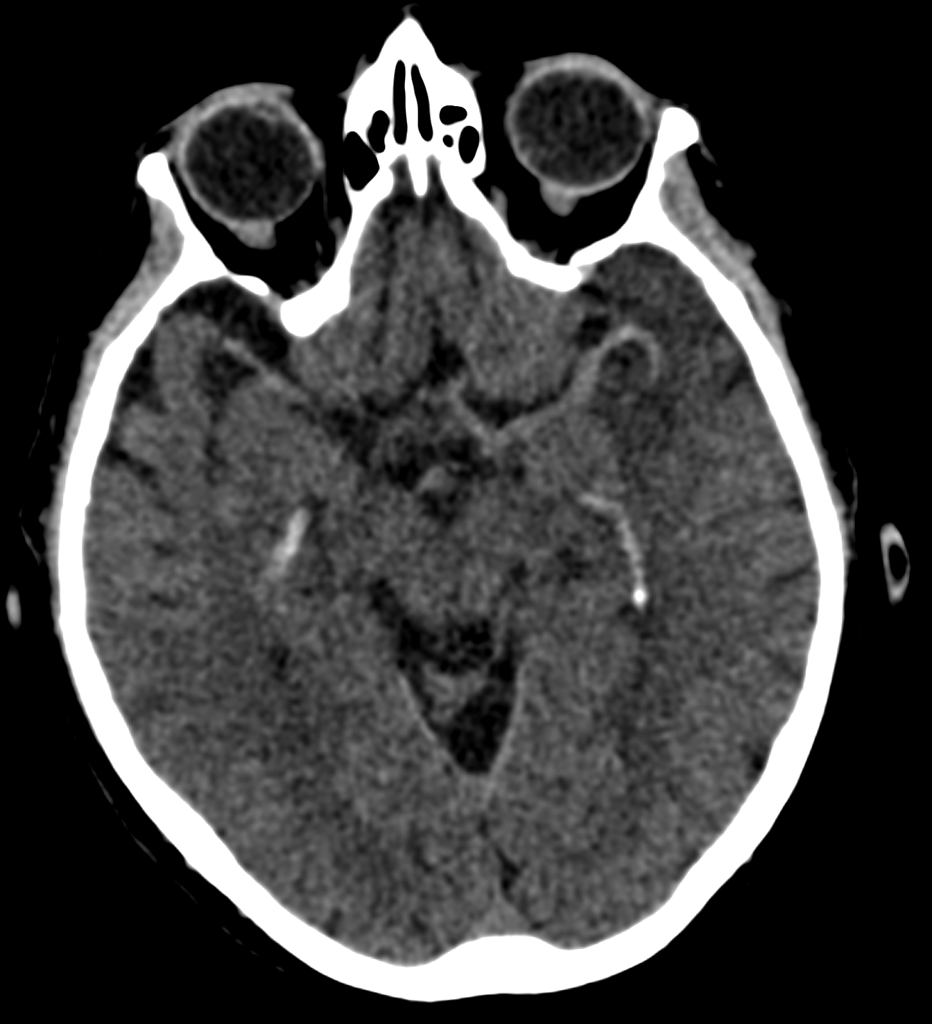
There is no abnormal intraparenchymal hyperdensity that is concerning for a parenchymal hemorrhage. Benign choroid plexus calcifications are present.
Compared to the brain parenchyma and the right middle cerebral artery, what is the density of the left middle cerebral artery?
The left middle cerebral artery is hyperdense compared to the remainder of the brain parenchyma, which suggests a thrombus within that artery.
The brain parenchyma is normal.
The left temporal pole is hypodense, which is indicative of cytotoxic edema in the setting of a suspected ischemic infarct.
View the full study if you'd like to take a look yourself.
Second Imaging Study
What is the next imaging study you will order?
The next step in the imaging algorithm should be CTA of the head and neck to evaluate for large-vessel occlusion. CTA and MRA perfusion technology can also help differentiate between irreversibly infarcted brain tissue and potentially salvageable tissue. This can help guide optimal treatment selection.
Well done. You were correct
Pertinent Imaging Observations
Click on the links below to view images from the study, and assess these key findings as best you can.
CTA head and neck
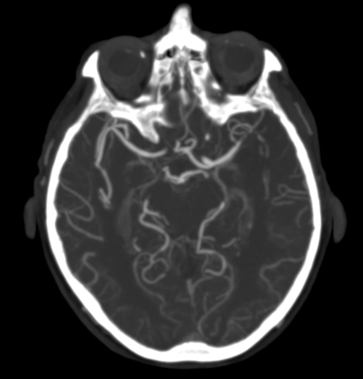
Is the M2 subdivision of the left MCA occluded in the CTA?
The M2 subdivision of the left MCA is occluded, as seen in the CTA.
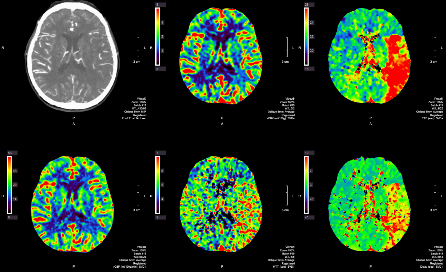
What does the increased TTP without significant changes in the CBV/CBF in the left temporal and parietal lobes suggest?
The increased TTP without significant changes in the CBV/CBF in the left temporal and parietal lobes suggest a salvageable penumbra in the left MCA distribution.
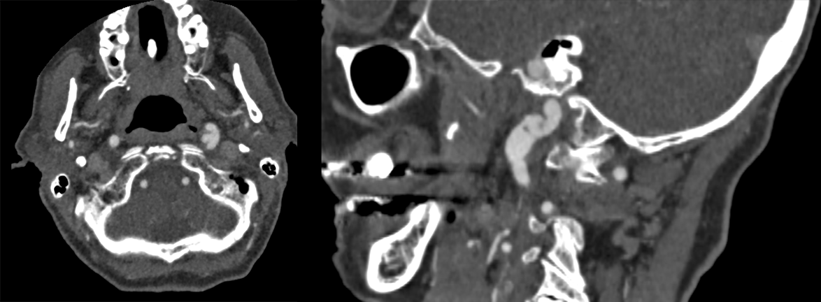
In the CTA of the neck, which of the following abnormalities is observed in an internal carotid artery?
There is a dissection flap and pseudoaneurysm in the left distal internal carotid artery.
View the full study if you'd like to like a look yourself
Third Imaging Study
What is the next imaging study you will order?
No further imaging is needed as the diagnosis is confirmed with the CTA.
What is your Diagnosis now that you have seen the imaging results?
This stroke may be amenable to intervention considering the presence of a salvageable penumbra on the CTA perfusion.
Current Acuity
Initially, you selected and we suggested acuity.
Has your concern for this patient changed?
The patient requires immediate management.
Assessment and Plan
Please provide your assessment and plan for this patient
The 85-year-old male patient with multiple risk factors, including hypertension, hyperlipidemia, type 2 diabetes mellitus, atrial fibrillation, and 60 pack-years of smoking, presents with an acute left M2 MCA stroke, as evidenced by the occlusion found on imaging and right-sided neurological deficits. There is a salvageable penumbra in the left MCA distribution. The stroke may have been secondary to the patient’s atrial fibrillation or the dissection flap and pseudoaneurysm in the left distal ICA. The patient should be admitted to the stroke unit and emergently evaluated by vascular neurology for thrombolytic therapy eligibility, considering the 2-hour onset of symptoms and the presence of a salvageable penumbra. The patient may require long term dual antiplatelet therapy due to the dissection flap and pseudoaneurysm. A stroke workup, including an echocardiogram and carotid ultrasound should be performed. Modifiable risk factors such as smoking hypertension, diabetes, hyperlipidemia, and atrial fibrillation, should be addressed prior to discharge.
Lessons Learned:
- Risk factors of ischemic stroke include older age, hypertension, hyperlipidemia, diabetes mellitus, atrial fibrillation, and smoking.
- Initial evaluation of ischemic stroke involves ensuring stability, assessing comorbidities, determining the etiology, and determining candidacy for reperfusion therapy.
- Brain imaging, including CT or MRI and neurovascular imaging with CT angiography or magnetic resonance angiography (MRA), plays a crucial role in diagnosing ischemic stroke, ruling out hemorrhage, and identifying vascular lesions responsible for the ischemic deficit. A non-contrast CT is particularly important for excluding hemorrhage in the initial phase.
- Treatment for eligible patients with intravenous thrombolysis should be initiated 4.5 prior to symptom onset of the time of their last known neurologic baseline. CTA and MRA perfusion technology can help differentiate between irreversibly infarcted brain tissue and potentially salvageable tissue, allowing for better selection of patients who may benefit from therapy.
- Eligible patients with acute ischemic stroke may receive intravenous thrombolytic therapy or mechanical thrombectomy, depending on the time frame and presence of a proximal large artery occlusion. Additional interventions associated with improved outcomes include antiplatelet therapy, prophylaxis for venous thromboembolism, and statin therapy.
Socioeconomic Factors: The connection between socioeconomic status and long-term stroke outcomes can be largely attributed to disparities in post-stroke care. Those with lower socioeconomic status frequently face inadequate access to essential healthcare and rehabilitation services.
That's the end of the module! Once you've reviewed the video(s), you can click here for another case challenge.
Next

