N10) Double vision after facial trauma
Review the Learning Outcomes, Hx, PE and Labs, and begin the module with your Provisional Diagnosis. Keep hitting "Next" to move through the module.
Learning Outcomes
- Articulate your relationship with the consulting diagnostic radiologists in the evaluation of a patient with orbital wall fracture.
- Review the DDx considerations in orbital wall fracture.
- Identify the spectrum of imaging findings in appropriate modalities for evaluating patients with orbital wall fractures.
History
Physical Exam
Labs
Provisional Diagnosis
Potential Acuity
What is your assessment of the likely acuity for this patient?
First Imaging Study
What is the first imaging study you will order?
Pertinent Imaging Observations
Click on the links below to view images from the study, and assess these key findings as best you can.
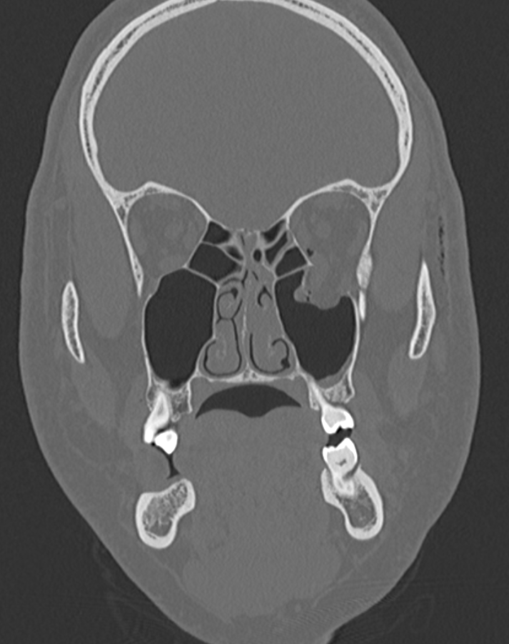
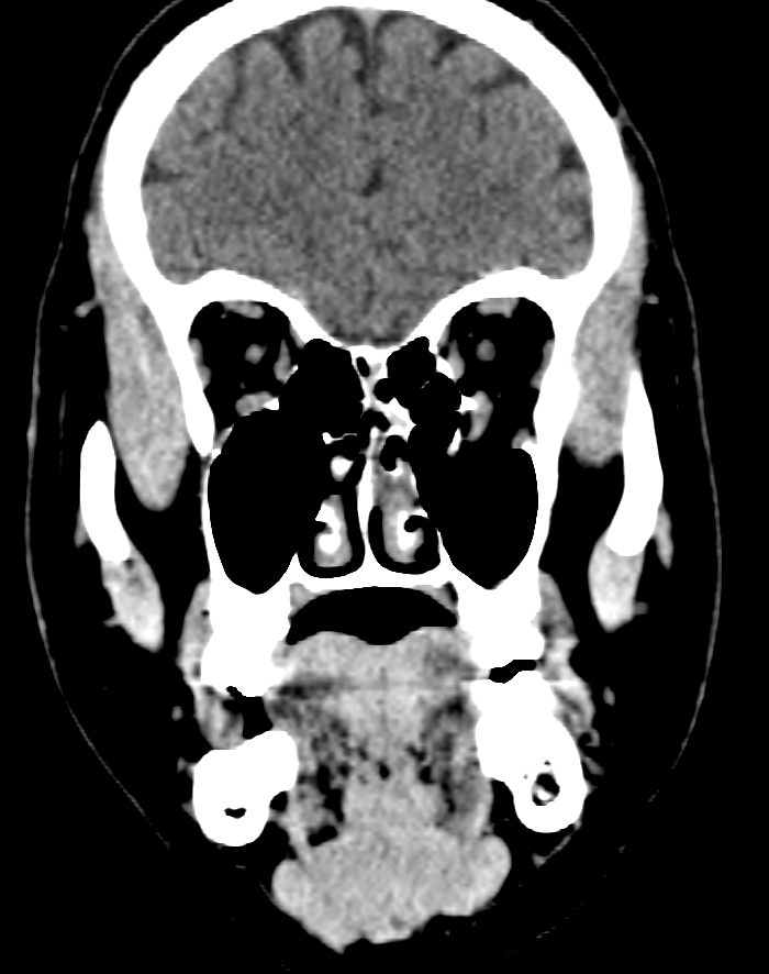
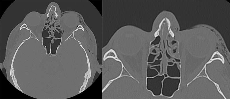
Head CT
There is an orbital wall fracture
There is herniation of the orbital adipose tissue into which of the following paranasal sinuses?
There is herniation of the inferior rectus muscle
There are other visualized fractures

Watch our video
Second Imaging Study
What is the next imaging study you will order?
What is your Diagnosis now that you have seen the imaging results?
Current Acuity
Initially, you selected and we suggested acuity.
Has your concern for this patient changed?
Assessment and Plan
Please provide your assessment and plan for this patient
Lessons Learned:
- Orbital wall blowout fractures often result from high-velocity blunt trauma to the eye causing herniation of the orbital contents into the maxillary sinus. CT is the modality of choice for making this diagnosis.
- Clinical features of orbital wall fractures include unilateral periorbital pain, edema, ecchymosis, and restricted ocular movement with or without diplopia if the rectus muscle is entrapped.
Socioeconomic Factors: Males between the ages of 16 and 35 are more likely to suffer from orbital wall fractures from trauma.
That's the end of the module! Once you've reviewed the video(s), you can click here for another case challenge.
Contributors:
Sean Kwak, MS2 - Content Contributor
Haoyu Wang - Content Contributor
Kevin Pierre, MD - Editor
Robbie Slater, MD - Supervising Editor
Bayar Batmunh, MS - Coordinator
