N1) Lucid interval following head injury
Review the Learning Outcomes, Hx, PE and Labs, and begin the module with your Provisional Diagnosis. Keep hitting "Next" to move through the module.
Learning Outcomes
- Articulate your relationship with the consulting diagnostic radiologists in the evaluation of a patient with altered mental status.
- Review the DDx considerations in a patient with altered mental status.
- Identify the spectrum of imaging findings in appropriate modalities for evaluating a patient with altered mental status.
History
Physical Exam
Labs
Provisional Diagnosis
Potential Acuity
What is your assessment of the likely acuity for this patient?
First Imaging Study
What is the first imaging study you will order?
Pertinent Imaging Observations
Click on the links below to view images from the study, and assess these key findings as best you can.
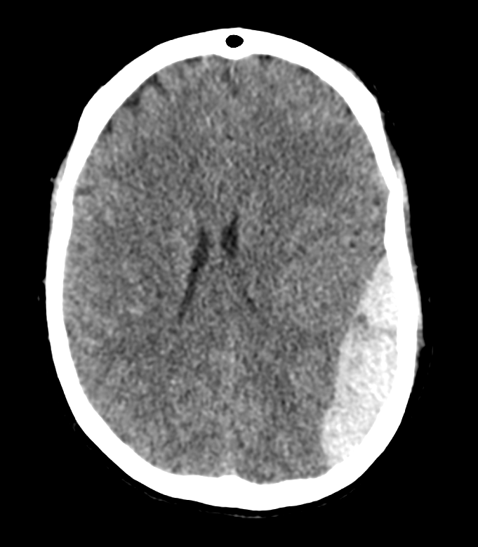
CT Head without IV Contrast
There is a subdural hematoma
There is midline shift.
There is a skull fracture.

Watch our video
Second Imaging Study
What is the next imaging study you will order?
What is your Diagnosis now that you have seen the imaging results?
Current Acuity
Initially, you selected and we suggested acuity.
Has your concern for this patient changed?
Assessment and Plan
Please provide your assessment and plan for this patient
Lessons Learned:
- Epidural hematoma often occurs secondary to head trauma and is typically a result of a rupture of the middle meningeal artery due to skull fracture. Blood accumulates between the skull and the dura mater, which leads to the characteristic lens-shaped hemorrhage, which respects the suture lines because the dura mater tightly adheres to the skull at the sutures. By contrast, depending on the location, a subdural hemorrhage classically crosses suture lines.
- Patients with epidural hematoma often experience a “lucid interval”, which is the period of alertness that occurs after losing consciousness and is followed by subsequent mental status decline. This clinical trajectory is explained by the accumulation of blood gradually increasing in size, which results in mass effect and neurological deficits.
- The first best diagnostic modality is the CT scan, which can detect intracranial bleeding as well as skull fracture.
Socioeconomic Factors:
- Mortality is highest in elderly patients and those with prolonged time between clinical intervention/surgery.
- Incidence of epidural hematoma is higher among males and adolescents.
- Uninsured patients, minority patients, and patients with more severe brain injuries face disproportionate treatment outcomes.
- Uninsured patients are less likely to be discharged to inpatient rehabilitation.
That's the end of the module! Once you've reviewed the video(s), you can click here for another case challenge.
Contributors:
Thomas Kent, MS2 - Content Contributor
Alexandria Hotop - Content Contributor
Kevin Pierre, MD - Editor
Robbie Slater, MD - Supervising Editor
Bayar Batmunh, MS - Coordinator
