Retake
M5) Foot pain after basketball injury
Review the Learning Outcomes, Hx, PE and Labs, and begin the module with your Provisional Diagnosis. Keep hitting "Next" to move through the module.
Learning Outcomes
- Articulate your relationship with the consulting diagnostic radiologists in the evaluation of a patient with foot pain.
- Review the DDx considerations in a patient with foot pain.
- Identify the spectrum of imaging findings in appropriate modalities for evaluating a patient with foot pain.
History
A 35-year-old professional basketball player comes into the clinic with left lateral foot pain. He states that 3 days ago he tripped while trying to pivot to the right and felt immediate foot pain. Afterwards there was bruising and swelling, and he unable to bear weight on the foot. The patient has been icing his foot and taking NSAIDS and Tylenol. His pain level 2/10 at rest and 10/10 when bearing weight.
Physical Exam
BP: 115/82, HR 55, RR 13, Temp 98.9, O2 saturation 100%. MSK: Edema and ecchymosis of left lateral foot. Point tenderness at the base of the fifth metatarsal.
Labs
None
Provisional Diagnosis
Select the Dx you believe is most appropriate
This patient most likely has a Jones fracture considering the lateral pain and point tenderness at the base of the fifth metatarsal after an injury with foot adduction and plantar flexion.
Well done. You were correct
Potential Acuity
What is your assessment of the likely acuity for this patient?
Well done. You were correct
The patient requires routine, but expedited workup.
First Imaging Study
What is the first imaging study you will order?
Foot radiographs are an appropriate initial imaging modality to evaluate for bony abnormalities in a patient with a suspected foot fracture. The Ottawa Ankle Rule suggest that imaging is appropriateness in this case.
Well done. You were correct
Pertinent Imaging Observations
Click on the links below to view images from the study, and assess these key findings as best you can.
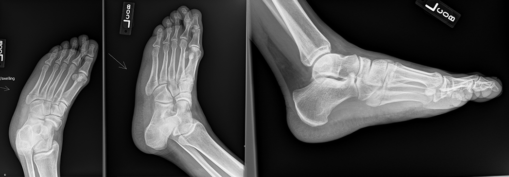
X-ray
The fracture is in which location of the fifth metatarsal?
There is a transverse fracture in the proximal metaphysis of the left fifth metatarsal, indicating it is a Jones fracture. A tuberosity fracture would likely be an avulsion fracture. A diaphyseal fracture would likely be a stress fracture or mid-shaft fracture.
The fracture is
There is no displacement of a fracture fragment to suggest displacement.
View the full study if you'd like to take a look yourself.
Second Imaging Study
What is the next imaging study you will order?
No further imaging is needed as the diagnosis was confirmed with radiography.
Well done. You were correct
What is your Diagnosis now that you have seen the imaging results?
This patient has a Jones fracture considering the lateral midfoot pain upon a pivoting injury and imaging findings.
Current Acuity
Initially, you selected and we suggested acuity.
Has your concern for this patient changed?
The patient requires routine, but expedited workup.
Assessment and Plan
Please provide your assessment and plan for this patient
This patient is a 35-year-old male presenting with a Jones fracture. Considering that the fracture is non-displaced, he may undergo nonoperative treatment with six to eight weeks in a nonweightbearing cast with analgestics as needed for pain. He is to return to clinic for follow-up radiographs to ensure continued alignment and healing. Considering the patient is a professional basketball player, a discussion should occur as to whether he would like to be evaluated for surgical intervention by orthopedic surgery to bear weight sooner.
Lessons Learned: - Jones fractures often occur as acute injuries involving plantar flexion and adduction of the foot. Patients may present with foot pain and swelling with tenderness at the base of the fifth metatarsal.
- Imaging shows a fracture line at the proximal metaphysis of the fifth metatarsal.
- The diagnosis should be suspected of injuries with lateral mid-foot pain and swelling.
- Conservative treatment may be undertaken for nondisplaced fractures. If the patient prefers to bear weight sooner, they may undergo evaluation for surgical intervention.
That's the end of the module! Once you've reviewed the video(s), you can click here for another case challenge.
Next

