M2) Shoulder pain after fall
Review the Learning Outcomes, Hx, PE and Labs, and begin the module with your Provisional Diagnosis. Keep hitting "Next" to move through the module.
Learning Outcomes
- Articulate your relationship with the consulting diagnostic radiologists in the evaluation of a patient with arm pain.
- Review the DDx considerations in a patient with arm pain.
- Identify the spectrum of imaging findings in appropriate modalities for evaluating a patient with arm pain.
History
Physical Exam
Labs
Provisional Diagnosis
Potential Acuity
What is your assessment of the likely acuity for this patient?
First Imaging Study
What is the first imaging study you will order?
Pertinent Imaging Observations
Click on the links below to view images from the study, and assess these key findings as best you can.
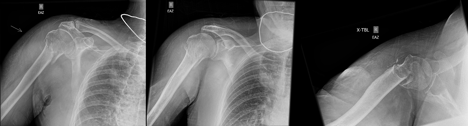
Right humerus X-ray
There is a joint dislocation.
There is a fracture.

Watch our video
Second Imaging Study
What is the next imaging study you will order?
What is your Diagnosis now that you have seen the imaging results?
Current Acuity
Initially, you selected and we suggested acuity.
Has your concern for this patient changed?
Assessment and Plan
Please provide your assessment and plan for this patient
Lessons Learned:
- Generally, when a proximal humeral fracture is suspected, a true AP view, axillary view, and scapular Y view (or Velpeau view if a scapular Y view is unobtainable). In this case, an adequate assessment was still possible with the provided views.
- It is important to classify proximal humerus fractures based on the type (transverse, oblique, spiral) and presence of displacement, impaction, or dislocation.
- The Neer classification is as follows: One-part: no displaced fragments; two-part: one displaced fragment; three-part: two-displaced fragments; four-part: 3+ displaced fragments + humeral head dislocation from the glenoid). Patients with Neer one-part fractures usually do well with closed, nonoperative management with a sling.
Socioeconomic Factors: - Proximal humerus fractures are most common in elderly female patients with osteoporosis. Elderly females most often face these fractures from low-energy mechanisms and face the higher mortality than younger males.
That's the end of the module! Once you've reviewed the video(s), you can click here for another case challenge.
Contributors:
Kevin Pierre, MD - Editor
Robbie Slater, MD - Supervising Editor
Bayar Batmunh, MS - Coordinator
