Retake
C15) Worsening hypoxia after hospitalization for severe burn injuries
Review the Learning Outcomes, Hx, PE and Labs, and begin the module with your Provisional Diagnosis. Keep hitting "Next" to move through the module.
Learning Outcomes
- Articulate your relationship with the consulting diagnostic radiologists in the evaluation of a patient with hypoxia.
- Review the DDx considerations in a patient with hypoxia.
- Identify the spectrum of imaging findings in appropriate modalities for evaluating a patient with hypoxia.
History
A 32-year-old female presented five days ago with 50% body surface area burns resulting in inhalational burn injury and bilateral lower extremity circumferential burns requiring escharotomies. She was intubated and subsequently had a tracheostomy implanted. She now is developing worsening hypoxia.
Physical Exam
BP: 123/79, HR 92, RR 104, Temp 36.7, O2 saturation 87%. CV: No pitting edema in legs. Normal S1/S2, no murmurs, rubs, or gallops. No jugular venous distension. Pulmonary: diffuse crackles.
Labs
WBC: 7.0 x 10^9L (nl: 4.5 x 10^9L - 11 x 10^9L);
PaO2: 50 mmHg;
FiO2: 0.4;
BNP: 15pg/mL (nL: <100).
Provisional Diagnosis
Select the Dx you believe is most appropriate
Acute respiratory distress syndrome is the most likely diagnosis considering the presence of a clinical insult (burns) with subsequent respiratory decline. Pneumonia is less likely considering the lack of fever and normal WBC count. Decompensated CHF is less likely considering the normal BNP and unremarkable cardiovascular physical exam.
Well done. You were correct
Potential Acuity
What is your assessment of the likely acuity for this patient?
Well done. You were correct
The patient requires urgent workup for her hypoxic respiratory failure.
First Imaging Study
What is the first imaging study you will order?
The chest X-ray is a good initial test for patients with respiratory decompensation. In this case, it may also detect a drainable pleural effusion or pneumothorax.
Well done. You were correct
Pertinent Imaging Observations
Click on the links below to view images from the study, and assess these key findings as best you can.
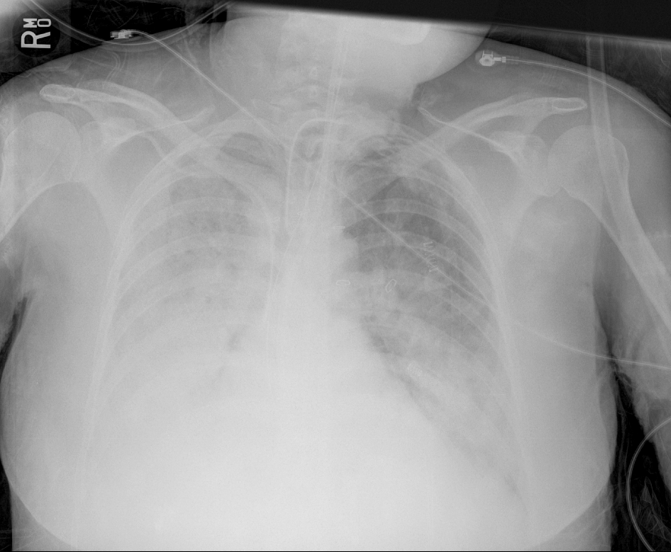
AP chest x-ray
What best describes the findings on the Chest X-ray?
The chest x-ray demonstrates diffuse, bilateral coalescent opacities representing alveolar infiltrates with a normal heart size, consistent with ARDS.
View the full study if you'd like to take a look yourself.
Second Imaging Study
What is the next imaging study you will order?
The clinical presentation and chest x-ray shows are consistent with ARDS. Therefore, no further imaging is required. A pleural ultrasound can be performed in place of CT to detect the presence of pleural effusions if there is a high clinical suspicion.
Well done. You were correct
What is your Diagnosis now that you have seen the imaging results?
The patient has moderate ARDS as the PaO2/FiO2 ratio is 125 (PAO2 ratios – Mild ARDS: 200-300, Moderate ARDS: 100-200, Severe ARDS: <100).
Current Acuity
Initially, you selected and we suggested acuity.
Has your concern for this patient changed?
This patient urgently requires respiratory support escalation prior to any further decompensation.
Assessment and Plan
Please provide your assessment and plan for this patient
Worsening hypoxic respiratory failure following severe burn injury. Imaging and labs are consistent with moderate ARDS. The patient will require increased respiratory support with judicious up-titration of FiO2 and PEEP.
Lessons Learned:
- ARDS results from an insult causing alveolar injury, leading to increased alveolar permeability and protein and cell rich exudate infiltration of the alveoli.
- Chest x-ray can show “white out” of one of both lungs secondary to diffuse, coalescent airspace opacities from alveolar filling.
- Unlike CHF which can have similar findings, the heart size is normal in ARDS.
- This case describes the exudative phase of ARDS. The proliferative phase occurs later and is often characterized by chronic pulmonary fibrosis.
Socioeconomic Factors: ARDS can be diagnosed with clinical findings, labs, and X-ray. A pleural ultrasound can be used in place of CT to detect the presence of pleural effusions if there is a high clinical suspicion. This reduces radiation exposure and is particularly useful where CT is not readily available.
That's the end of the module! Once you've reviewed the video(s), you can click here for another case challenge.
Next

