A19) RLQ abdominal pain
Review the Learning Outcomes, Hx, PE and Labs, and begin the module with your Provisional Diagnosis. Keep hitting "Next" to move through the module.
Learning Outcomes
- Articulate your relationship with consulting diagnostic radiologists, in RLQ abdominal pain.
- Review the DDx considerations in RLQ abdominal pain scenarios.
- Identify the spectrum of imaging findings in appropriate modalities for evaluating RLQ pain.
History
Physical Exam
Labs
Provisional Diagnosis
Potential Acuity
What is your assessment of the likely acuity for this patient?
First Imaging Study
What is the first imaging study you will order?
Pertinent Imaging Observations
Click on the links below to view images from the study, and assess these key findings as best you can.
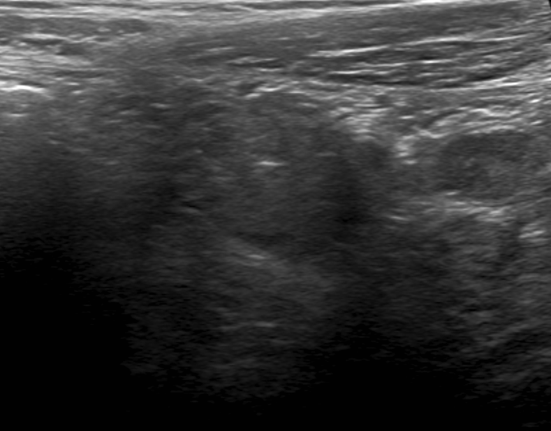
Ultrasound Observations
The appendix is clearly visible on the ultrasound.

Watch our video
Second Imaging Study
What is the next imaging study you will order?
Pertinent Imaging Observations
Click on the links below to view images from the study, and assess these key findings as best you can.
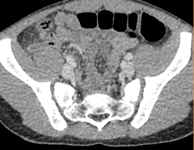
CT Abdomen and Pelvis
The appendix is dilated and fluid filled.
Third Imaging Study
What is the next imaging study you will order?
What is your Diagnosis now that you have seen the imaging results?
Current Acuity
Initially, you selected and we suggested acuity.
Has your concern for this patient changed?
Assessment and Plan
Please provide your assessment and plan for this patient
Lessons Learned
Socioeconomic Factors: The diagnosis of appendicitis can often be made by ultrasound, especially in the pediatric population. It would be lower cost and there is no radiation exposure. However, the appendix may be obscured by bowel gas and a CT would be necessary if clinical suspicion remains high.
That's the end of the module! Once you've reviewed the video(s), you can click here for another case challenge.
