Retake
A28) Hematuria, flank pain, and weight loss
Review the Learning Outcomes, Hx, PE and Labs, and begin the module with your Provisional Diagnosis. Keep hitting "Next" to move through the module.
Learning Outcomes
- Articulate your relationship with the consulting diagnostic radiologists in the evaluation of a patient with hematuria.
- Review the DDx considerations for a patient with hematuria.
- Identify the spectrum of imaging findings in appropriate modalities for evaluating a patient with hematuria.
History
A 70-year-old male visits his primary care physician due to intermittent episodes of hematuria and intermittent left flank pain. He has unintentionally lost 25 pounds over the last three months. He also has an 80-pack year smoking history and denies taking any new medications.
Physical Exam
BP: 130/90, HR 70, RR 16, Temp 37º C, O2 saturation 98%, BMI: 34.
Abdominal exam: Minimal left flank tenderness.
Labs
Hgb: 10.2 g/dL (normal range for males: 13.5 to 17.5 g/dL).
Urinalysis: microscopic hematuria.
Provisional Diagnosis
Select the Dx you believe is most appropriate
The patient most likely has renal cell carcinoma, given his age, significant smoking history, and the presence of weight loss, hematuria, and left flank pain.
Well done. You were correct
Potential Acuity
What is your assessment of the likely acuity for this patient?
Well done. You were correct
The patient requires routine, but expedited workup.
First Imaging Study
What is the first imaging study you will order?
Contrast-enhanced CT scans are highly effective in characterizing renal cell tumors, accurately identifying their size, location, and characteristics, and detecting any invasion of nearby structures. Notably, they are highly sensitive and specific in detecting renal cell carcinoma.
Well done. You were correct
Pertinent Imaging Observations
Click on the links below to view images from the study, and assess these key findings as best you can.
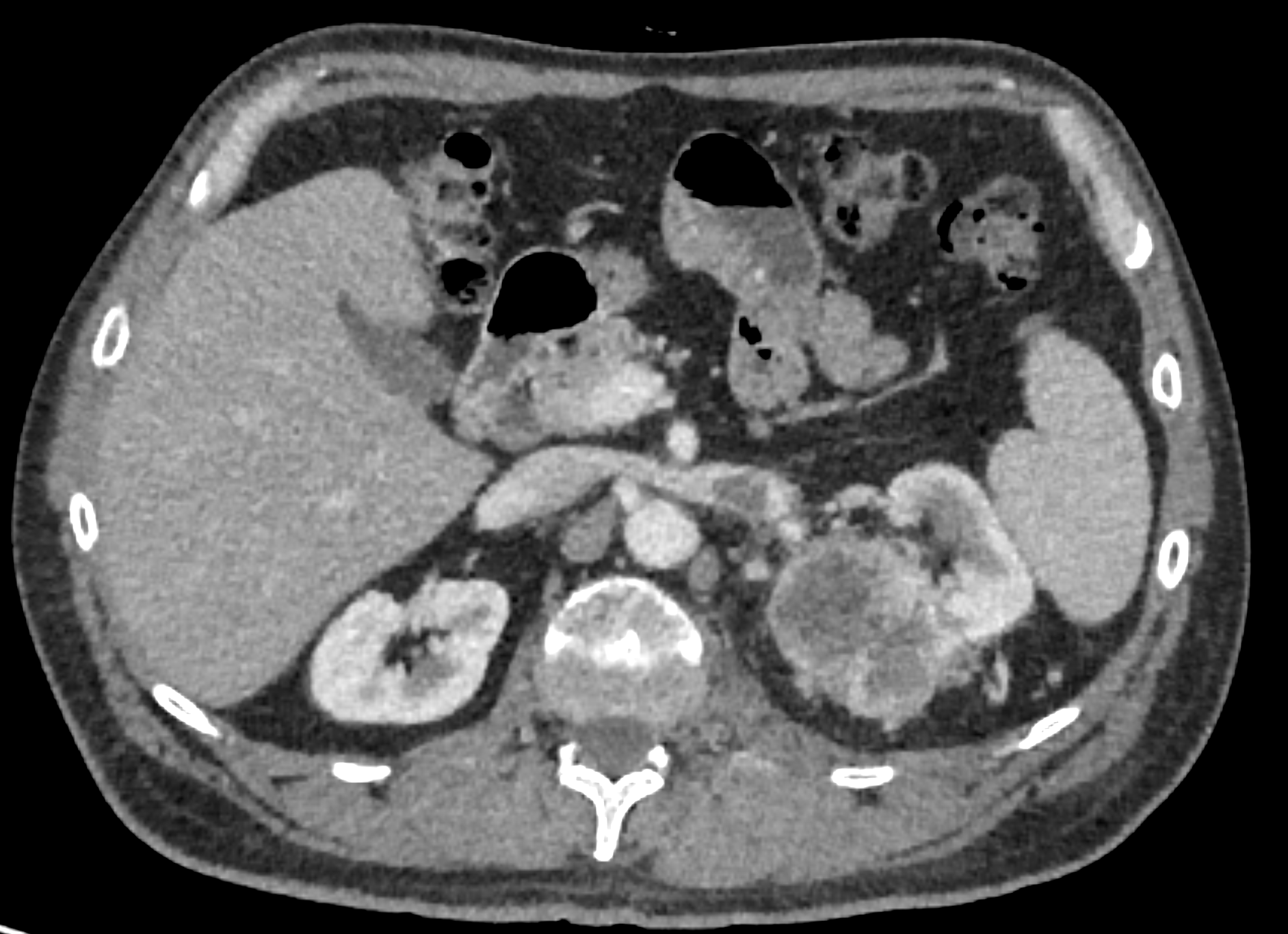
CT Abdomen and Pelvis
What is present in the left kidney?
There is a large, centrally necrotic lesion with a thick, irregular, and enhancing wall and internal septations. This lesion can also be classified as a malignant Bosniak IV renal cyst, and is therefore most likely malignant. These features are highly suggestive of renal cell carcinoma.
There is evidence of invasion into which nearby structure?
The left renal vein exhibits areas of hypoattenuation, possibly representing a tumor thrombus. Renal cell carcinoma is known to invade the renal vein.
View the full study if you'd like to take a look yourself.
Second Imaging Study
What is the next imaging study you will order?
The diagnosis is confirmed with CT. However, a spinal MRI or PET scan may be required for staging.
Well done. You were correct
What is your Diagnosis now that you have seen the imaging results?
This patient has renal cell carcinoma given the left renal mass that is invading into nearby structures.
Current Acuity
Initially, you selected and we suggested acuity.
Has your concern for this patient changed?
The patient requires routine but expedited workup.
Assessment and Plan
Please provide your assessment and plan for this patient
The patient is a 70-year-old male presenting with a left renal mass concerning for renal cell carcinoma with invasion into the left renal vein and infiltration into the psoas, paraspinous muscles, and L2 vertebra, extending into the spinal canal from L1-L3. The patient should be referred to a urologist for further staging and management. Possible treatment options include radical or partial nephrectomy, arterial embolization, immunotherapy, targeted therapy (e.g. VEGF or mTOR inhibitors), or radiation therapy, depending on the stage of the disease and the patient's overall health. The patient should also receive smoking cessation counseling.
Lessons Learned:
- Renal cell carcinoma (RCC) is the most common type of kidney cancer and is more common in older men. Major risk factors for RCC include smoking, obesity, hypertension, and a family history of the disease.
- Common symptoms of RCC include hematuria, flank pain, an abdominal mass, and unintentional weight loss. However, many RCCs are asymptomatic and discovered incidentally on imaging studies performed for other reasons.
- CT scan with IV contrast is the imaging modality of choice for diagnosing RCC, as it can reveal the tumor, invasion into nearby structures, and metastatic disease.
- Early diagnosis and treatment of RCC are crucial for improving patient outcomes. Treatment options include surgery (partial or radical nephrectomy), arterial embolization, immunotherapy, targeted therapy, and radiation therapy.
That's the end of the module! Once you've reviewed the video(s), you can click here for another case challenge.
Next

