Retake
A7) Flank pain and vomiting after cystitis
Review the Learning Outcomes, Hx, PE and Labs, and begin the module with your Provisional Diagnosis. Keep hitting "Next" to move through the module.
Learning Outcomes
- Articulate your relationship with the consulting diagnostic radiologists in the evaluation of a patient with flank pain.
- Identify the spectrum of imaging findings in appropriate modalities for evaluating a patient with flank pain.
History
A 27-year-old homeless female with a history of uncontrolled diabetes and multiple sclerosis requiring rituximab for a recent flare-up presents to the ED with flank pain. She is somnolent but arousable. She was diagnosed with cystitis four days ago and has missed several antibiotic doses. Her dysuria, suprapubic pain, urinary urgency and frequency have not improved. She endorses new onset nausea, vomiting, and flank pain.
Physical Exam
BP: 87/51, HR 114, RR 21, Temp 40.1C, O2 saturation 92%.
Abdominal: Severe costovertebral angle (CVA) bilaterally and suprapubic tenderness. Abdomen otherwise soft and non-peritonitic.
Labs
WBC: 23 x 10^9/L with left shift. Lactate >4 mmol/L.
Urinalysis with microscopy: 15wbc/hpf, 5 rbc/hpf, and WBC casts.
Urinalysis gram stain: gram-negative bacilli.
A1C: 10.4%
Provisional Diagnosis
Select the Dx you believe is most appropriate
An ascending infection from the cystitis resulting in pyelonephritis is most likely in this case considering the recent cystitis and new onset flank pain, nausea, and vomiting.
Well done. You were correct
Potential Acuity
What is your assessment of the likely acuity for this patient?
Well done. You were correct
The patient meets criteria for sepsis and requires admission and further workup.
First Imaging Study
What is the first imaging study you will order?
The diagnosis of pyelonephritis can be made clinically. However, the patient’s uncontrolled diabetes and immunosuppressed status due to rituximab use significantly increases her risk for complications like an abscess and papillary necrosis. Therefore, imaging is appropriate in this case. A non-contrast CT can assess for renal stones. Contrast enhanced CT better allows for visualization of the renal parenchyma and associated complications.
Well done. You were correct
Pertinent Imaging Observations
Click on the links below to view images from the study, and assess these key findings as best you can.
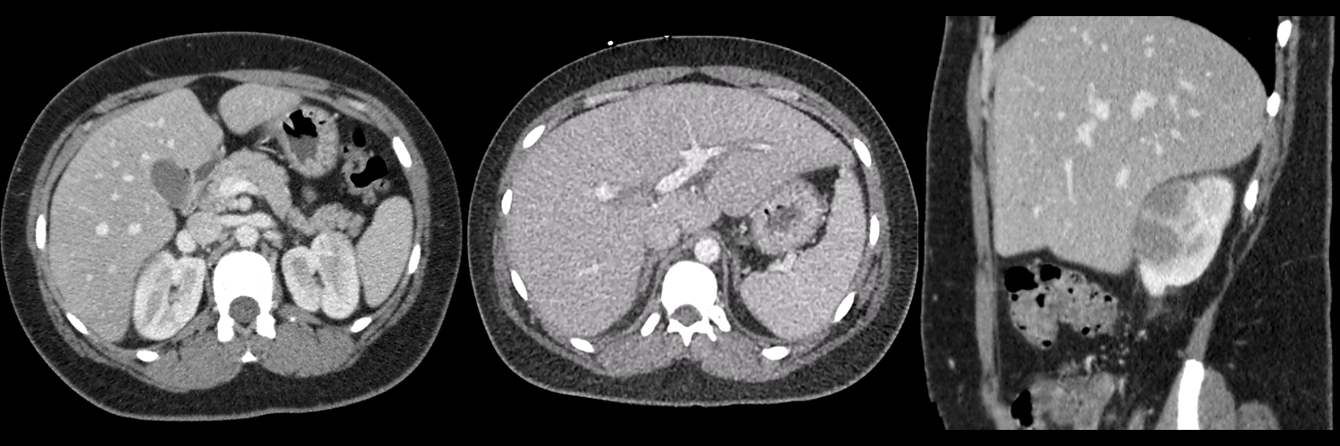
CT scan with and without contrast
There are wedge-like areas of decreased enhancement in the kidneys.
There are wedge shaped areas of decreased enhancement in both kidneys. These likely represent pyelonephritis. These are less likely to represent renal infarcts as the periphery of the renal cortex is involved.
Which of the following is present in the right kidney?
The fluid collection in the right kidney likely represents a corticomedullary abscess. A perinephric abscess would be adjacent to the renal parenchyma.
There is perinephric stranding.
There is perinephric stranding, most significant in the right kidney, secondary to the pyelonephritis.
View the full study if you'd like to take a look yourself.
Second Imaging Study
What is the next imaging study you will order?
The patient will require serial imaging to follow evolution of the abscess.
Well done. You were correct
Pertinent Imaging Observations
Click on the links below to view images from the study, and assess these key findings as best you can.
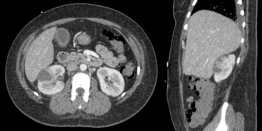
CT abdomen with contrast
The subsequent CT scan indicates the presence of:
The evolution of previous lesions is now apparent. These lesions exhibit hypoenhancing central components accompanied by peripheral enhancement, which confirms that they are multifocal abscesses.
View the full study if you'd like to like a look yourself
Third Imaging Study
What is the next imaging study you will order?
The patient will require serial imaging to follow evolution of the abscess.
What is your Diagnosis now that you have seen the imaging results?
Current Acuity
Initially, you selected and we suggested acuity.
Has your concern for this patient changed?
The patient requires admission and prompt treatment.
Assessment and Plan
Please provide your assessment and plan for this patient
The patient is a 27-year-old female presenting with sepsis secondary to acute pyelonephritis with a corticomedullary abscess. She will require admission for IV antibiotics and IV fluids. We will obtain urine and blood cultures to later narrow our antibiotic regimen. We will obtain another CT scan in a short interval to evaluate for change and progression.
Lessons Learned:
- Pyelonephritis can occur secondary to ascending infection from cystitis. This ascending infection and complications are more likely in patients with diabetes, immunocompromise, indwelling Foley catheters, and urinary tract abnormalities including renal calculi, prostatic hyperplasia, and strictured urethra.
Socioeconomic Factors: Women from non-Western countries and low socioeconomic status are at higher risk for pyelonephritis.
That's the end of the module! Once you've reviewed the video(s), you can click here for another case challenge.
Next

