A31) Epigastric pain
Review the Learning Outcomes, Hx, PE and Labs, and begin the module with your Provisional Diagnosis. Keep hitting "Next" to move through the module.
Learning Outcomes
- Articulate your relationship with the consulting diagnostic radiologists in the evaluation of a patient with epigastric pain.
- Review the DDx considerations in a patient with epigastric pain.
- Identify the spectrum of imaging findings in appropriate modalities for evaluating a patient with epigastric pain.
History
Physical Exam
Labs
Provisional Diagnosis
Potential Acuity
What is your assessment of the likely acuity for this patient?
First Imaging Study
What is the first imaging study you will order?
Pertinent Imaging Observations
Click on the links below to view images from the study, and assess these key findings as best you can.
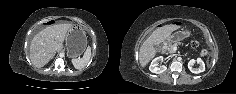
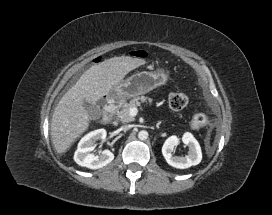
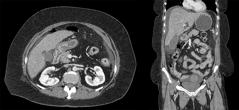
CT chest, abdomen, and pelvis with IV contrast
There is free intraperitoneal air
There is a locule of air adjacent to the duodenum
The duodenum is normal
There is free intraperitoneal fluid

Watch our video
Second Imaging Study
What is the next imaging study you will order?
What is your Diagnosis now that you have seen the imaging results?
Current Acuity
Initially, you selected and we suggested acuity.
Has your concern for this patient changed?
Assessment and Plan
Please provide your assessment and plan for this patient
Lessons Learned:
- Perforation is a complication of peptic ulcer disease. The most common complication is upper GI tract hemorrhage.
- A perforated ulcer should be suspected if the patient presents with acute worsening of pain with peritoneal signs. In this case, a CT scan with IV contrast should be performed which often shows pneumoperitoneum and discontinuity in the duodenal wall or stomach with adjacent air locules.
- Otherwise, if an non-perforated ulcer is suspected, EGD or fluoroscopy are the appropriate diagnostic methods.
Socioeconomic Factors: Management of risk factors from a primary care standpoint can significantly reduce risk of ulcer formation and subsequent perforation. For example, patients who smoke should be provided with resources for smoking cessation and patients with chronic pain should be educated on dangers of chronic NSAID use. Patients with peptic ulcer disease should also be educated on signs of ulcer perforation. Studies have shown that patients who present earlier and undergo immediate management had better outcomes than those that presented more than 24 hours after symptom onset.
That's the end of the module! Once you've reviewed the video(s), you can click here for another case challenge.
Contributors:
Kevin Pierre, MD - Editor
Robbie Slater, MD - Supervising Editor
Bayar Batmunh, MS - Coordinator
