Retake
A21) RUQ abdominal pain, nausea, and vomiting
Review the Learning Outcomes, Hx, PE and Labs, and begin the module with your Provisional Diagnosis. Keep hitting "Next" to move through the module.
Learning Outcomes
- Articulate your relationship with the consulting diagnostic radiologists in the evaluation of a patient with right upper quadrant pain.
- Review the DDx considerations in a patient with right upper quadrant pain.
- Identify the spectrum of imaging findings in appropriate modalities for evaluating a patient with right upper quadrant pain.
History
An 80-year-old male arrived at the emergency department after experiencing severe, cramping pain in his right upper quadrant over the past two days. The pain, radiating towards his right scapula, was accompanied by symptoms of nausea, vomiting, low-grade fever, and loss of appetite. He notes that a similar discomfort, particularly after consuming fatty foods, has occurred intermittently over the past three months. The patient received analgesics during transport by EMS.
Physical Exam
Blood Pressure: 110/70 mmHg; Heart Rate: 100 bpm; Respiratory Rate: 20 breaths/min; Temperature: 38.9ºC (102F); Oxygen Saturation: 97% on room air. Abdominal Exam: Right upper quadrant tenderness to palpation, but Murphy's sign is negative. Voluntary guarding present. No other palpable masses or organomegaly noted.
Labs
White Blood Cell (WBC) Count: 13,000/µL (normal: 4,500-11,000/µL).
Alanine aminotransferase (ALT): 22 U/L (normal: 7-55 U/L),
Aspartate aminotransferase (AST): 28 U/L (normal: 8-48 U/L),
Alkaline Phosphatase (ALP): 70 U/L (normal: 40-129 U/L),
Total Bilirubin: 0.9 mg/dL (normal: 0.3-1.2 mg/dL),
Direct Bilirubin: 0.2 mg/dL (normal: 0.0-0.3 mg/dL).
Provisional Diagnosis
Select the Dx you believe is most appropriate
Considering the history of postprandial abdominal pain, current presentation of severe right upper quadrant pain radiating to the right scapula (secondary to irritation of the right phrenic nerve), associated symptoms, and laboratory findings including fever and leukocytosis, the most probable diagnosis is acute cholecystitis. Although acute cholecystitis typically presents with a positive Murphy's sign, it is likely that the sign is negative in this instance due to the analgesics administered en route. Given the normal liver function tests, conditions such as ascending cholangitis, and alcoholic hepatitis are less likely.
Well done. You were correct
Potential Acuity
What is your assessment of the likely acuity for this patient?
Well done. You were correct
The patient requires urgent workup and management.
First Imaging Study
What is the first imaging study you will order?
A right upper quadrant ultrasound is the best initial imaging modality to evaluate a patient with right upper quadrant pain and suspected biliary disease.
Well done. You were correct
Pertinent Imaging Observations
Click on the links below to view images from the study, and assess these key findings as best you can.
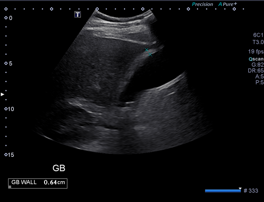
RUQ US
There is gallbladder wall thickening.
The thickness of a healthy gallbladder wall should not exceed 3 mm. In this patient, the gallbladder wall is thickened to 6.4 mm.
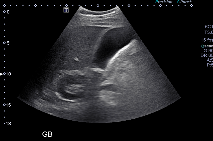
Which sonographic phenomenon suggests the presence of a gallstone?
Acoustic shadowing is a sonographic phenomenon observed when the ultrasound waves encounter a solid or high-density object, such as a gallstone. This results in a reduction of the sound wave signal distal to the object, creating a 'shadow'. In the patient's case, the presence of a lesion that produces acoustic shadowing strongly suggests the presence of a gallstone located at the neck of the gallbladder, irrespective of its composition.
View the full study if you'd like to take a look yourself.
Second Imaging Study
What is the next imaging study you will order?
A HIDA scan should be considered in indeterminate cases of cholecystitis where a sonographic Murphy’s sign is negative.
Well done. You were correct
Pertinent Imaging Observations
Click on the links below to view images from the study, and assess these key findings as best you can.
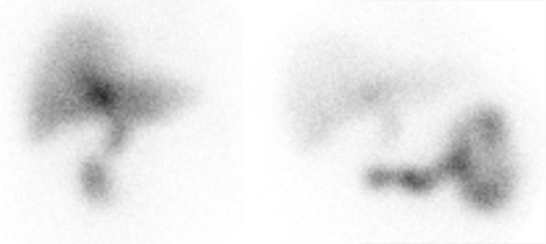
HIDA scan
Has the radiotracer been taken up by the gallbladder?
In this case, there is an absence of radiotracer uptake within the gallbladder, which is strongly indicative of acute cholecystitis. This is because the obstruction by the stone prevents normal uptake of radiotracer by the gallbladder.
There is abnormally increased uptake of radiotracer in the liver.
There is the presence of a "rim sign" - characterized by increased uptake of the radiotracer along the liver parenchyma adjacent to the gallbladder fossa. This sign implies the likely presence of gangrenous cholecystitis. The mechanism behind the rim sign involves severe inflammation leading to gallbladder wall ischemia and necrosis. The liver parenchyma adjacent to the necrotic gallbladder becomes hyperemic and has increased blood flow, leading to increased uptake of the radiotracer.
View the full study if you'd like to like a look yourself
Third Imaging Study
What is the next imaging study you will order?
No further imaging is needed.
What is your Diagnosis now that you have seen the imaging results?
The patient’s presentation, physical exam findings and imaging are highly suggestive of acute gangrenous cholecystitis secondary to cholelithiasis.
Current Acuity
Initially, you selected and we suggested acuity.
Has your concern for this patient changed?
This patient requires an emergent evaluation and management plan. Gangrenous cholecystitis is a serious complication that can lead to gallbladder perforation and subsequent sepsis if not promptly managed.
Assessment and Plan
Please provide your assessment and plan for this patient
The patient, an 80-year-old male, presents with right upper quadrant pain, fever, nausea, and vomiting. Based on the clinical presentation and imaging findings, a diagnosis of severe acute cholecystitis is highly likely. The hepatobiliary scintigraphy findings, particularly the presence of the "rim sign," suggest a 30-40% likelihood of gangrenous cholecystitis, which is a serious and potentially life-threatening condition. General surgery should be urgently consulted for further evaluation and management and a possible laparoscopic cholecystectomy. Concurrently, IV fluids, analgesics, antiemetics, and broad-spectrum antibiotics should be administered.
Lessons Learned:
- Acute cholecystitis, commonly a complication of gallstone disease, occurs due to cystic duct obstruction and potential bile infection, usually in patients with a history of symptomatic gallstones.
- Patients typically present with prolonged, severe pain in the right upper quadrant or epigastric region, which occurs after consuming fatty foods. This pain often radiates to the right shoulder or back and is accompanied by symptoms such as nausea, vomiting, and anorexia.
- A negative Murphy's sign does not rule out a diagnosis of cholecystitis, particularly after the administration of analgesics.
- Gangrenous cholecystitis with perforation may occur in advanced cases. Additional complications can include pericholecystic abscess, pyogenic liver abscess, and hemorrhagic cholecystitis.
- The most appropriate initial imaging modality is a right upper quadrant ultrasound, which can reveal an obstructing stone, gallbladder wall thickening, and pericholecystic fluid.
- A HIDA scan is useful in evaluating cases of cholecystitis that are indeterminate, where a sonographic Murphy's sign is negative, or when the ultrasound results are inconclusive. The “rim sign” suggests the presence of gangrenous cholecystitis.
That's the end of the module! Once you've reviewed the video(s), you can click here for another case challenge.
Next

