Retake
A20) Acute-onset abdominal pain and bilious vomiting
Review the Learning Outcomes, Hx, PE and Labs, and begin the module with your Provisional Diagnosis. Keep hitting "Next" to move through the module.
Learning Outcomes
- Articulate your relationship with the consulting diagnostic radiologists in the evaluation of a patient with right lower quadrant pain.
- Review the DDx considerations in a patient with right lower quadrant pain.
- Identify the spectrum of imaging findings in appropriate modalities for evaluating a patient with right lower quadrant pain.
History
A 28-year-old female presents with acute onset of nausea, bilious vomiting and abdominal pain that has worsened over the past day. She also complains of abdominal distension. She has not passed flatus or stool since the onset of symptoms. She has no history of abdominal surgeries.
Physical Exam
BP: 115/70, HR 99, RR 18, Temp 98.6, O2 saturation 97%.
General: Mild distress.
Abdominal: High-pitched bowel sounds. Diffuse tenderness to palpation. Abdomen tympanic to percussion. No rebound tenderness or guarding.
Labs
None
Provisional Diagnosis
Select the Dx you believe is most appropriate
Given the patient's severe abdominal pain, distension, and obstipation, the presentation is highly suggestive of a bowel obstruction.
Well done. You were correct
Potential Acuity
What is your assessment of the likely acuity for this patient?
Well done. You were correct
In this patient, there are no signs of bowel ischemia, such as rebound tenderness, rigidity, or guarding, which would indicate a need for emergent surgery. Furthermore, the patient is hemodynamically stable. However, due to the concern for bowel obstruction, urgent workup and management is required to prevent potential vascular compromise, bowel ischemia, and perforation.
First Imaging Study
What is the first imaging study you will order?
In patients with suspected high-grade small bowel obstruction (SBO), the most appropriate initial imaging modality is a CT scan of the abdomen and pelvis. CT scans are accurate in identifying the presence and often, the etiology of obstruction, and detecting the presence of complications, such as strangulation and ischemia. Additionally, CT scans can guide patient management, helping triaging them into operative or nonoperative treatment groups based on the findings.
Well done. You were correct
Pertinent Imaging Observations
Click on the links below to view images from the study, and assess these key findings as best you can.
CT Abdomen
What best describes the findings on the Abdominal CT?
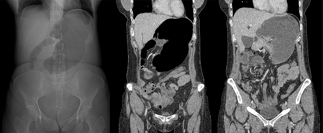
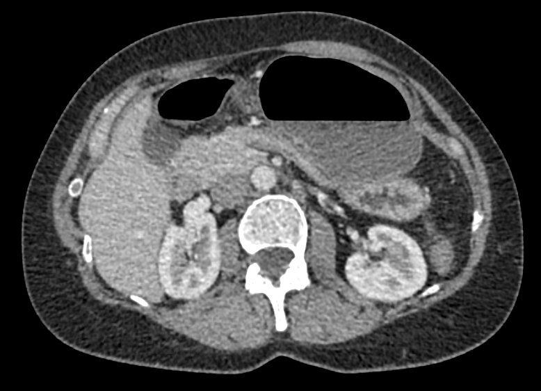
The presence of a dilated cecum in the left upper quadrant, along with dilated small bowel and the presence of a "coffee-bean" sign pointing to the left upper quadrant, indicates the presence of a cecal volvulus. The "whirl sign," demonstrating twisting of the mesentery with its vascular pedicle, is observed.
There is intraabdominal free air.
There is no evidence of sequelae related to vascular compromise, such as bowel ischemia characterized by pneumatosis, or perforation indicated by pneumoperitoneum.
View the full study if you'd like to take a look yourself.
Second Imaging Study
What is the next imaging study you will order?
No additional imaging is necessary as the diagnosis of cecal volvulus has been confirmed with the abdominal CT scan.
Well done. You were correct
What is your Diagnosis now that you have seen the imaging results?
This patient’s presentation is most consistent with a cecal volvulus.
Current Acuity
Initially, you selected and we suggested acuity.
Has your concern for this patient changed?
The patient requires urgent workup and management.
Assessment and Plan
Please provide your assessment and plan for this patient
This is a 28-year-old hemodynamically stable female presenting with severe abdominal pain, distension, and obstipation. A CT scan confirms the diagnosis of cecal volvulus. The patient should be made NPO, undergo nasogastric decompression, and receive analgesics. General surgery should be consulted for further management. Given her stable condition without bowel compromise, the management plan is likely to involve detorsion followed by either a right colectomy or an ileocolic resection.
Lessons Learned:
- Volvulus accounts for 10-15% of intestinal obstructions in adults in the US, making it the third most common cause.
- Although rare, cecal volvulus is more frequently seen in middle-aged females.
- The pathophysiology involves the torsion of the colonic mesenteric vascular pedicle, resulting in vascular compromise that can progress to ischemia and bowel strangulation.
- Imaging studies may reveal a dilated cecum with a characteristic appearance known as the "coffee-bean" sign, pointing toward the left upper quadrant. The presence of the "whirl sign," demonstrating twisting of the mesentery with its vascular pedicle, is also indicative of volvulus.
- It is crucial to assess for potential complications of vascular compromise on imaging, such as bowel ischemia characterized by pneumatosis, or perforation demonstrated by pneumoperitoneum.
- The management of cecal volvulus varies significantly and is influenced by the patient's hemodynamic stability and the presence of bowel compromise.
That's the end of the module! Once you've reviewed the video(s), you can click here for another case challenge.
Next

