Case Notes
History
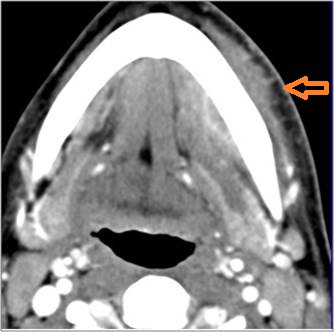
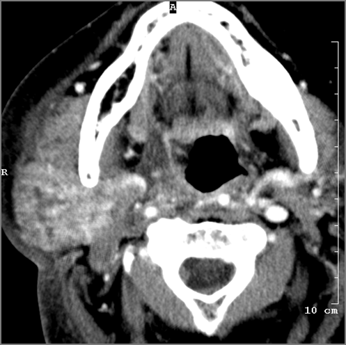
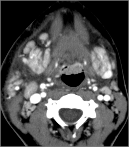
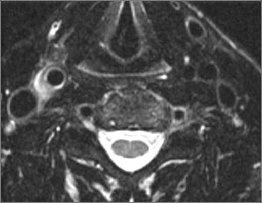
32-year old female with fever and severe throat pain and shortness of breath for two days. Clinical suspicion of pharyngitis and retropharyngeal abscess.Exam
Prior Study
Dicom
Findings
| General | Correct Answer | Your Answer |
|---|---|---|
|
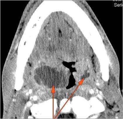
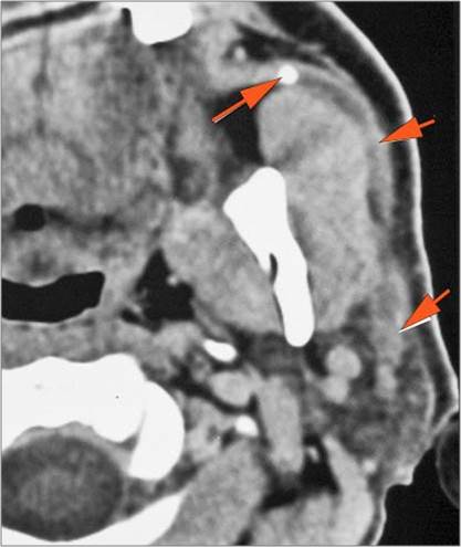
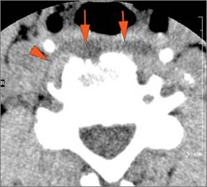
There is general or localized soft tissue swelling of the superficial or deep neck soft tissues. |
Yes | NA |
|
There is gas in the soft tissues. |
No | NA |
|
There is a foreign body separate from or in areas of soft tissue swelling. |
No | NA |
| Nasopharynx | Correct Answer | Your Answer |
|---|---|---|
|
There is excessive enhancement or thickening of the mucosa or hypertrophy of the lymphoid tissue in the nasopharynx. |
Yes | NA |
|
There is evidence of an abscess within the lymphoid tissue of the nasopharynx. |
No | NA |
|
There is edema within the fat of the adjacent parapharyngeal space. |
Yes | NA |
|
There is an abscess within the fat of the adjacent parapharyngeal space. |
No | NA |
|
There is edema within the fat of the adjacent retropharyngeal space. |
No | NA |
|
There is an abscess within the fat of the adjacent retropharyngeal space. |
No | NA |
| Oropharynx | Correct Answer | Your Answer |
|---|---|---|
|
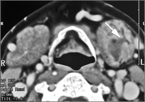
There is excessive enhancement or thickening of the mucosa or hypertrophy of the palatine or lingual tonsillar tissue or the lymphoid tissue along the glossotonsillar sulci and posterior pharyngeal wall. |
Yes | NA |
|
There is abscess relatively centrally within the lymphoid tissue of the palatine tonsil. |
No | NA |
|
There is abscess at the periphery of the lymphoid tissue of the palatine tonsil within the potential peritonsillar spaceof the palatine tonsil within the potential peritonsillar space |
No | NA |
|
There is edema/abscess within the fat of the adjacent parapharyngeal and retropharyngeal spaces. |
Yes | NA |
| Retropharyngeal Lymph Nodes | Correct Answer | Your Answer |
|---|---|---|
|
There is reactive retropharyngeal lymphadenopathy. |
No | NA |
|
There is suppurative retropharyngeal lymphadenopathy. |
No | NA |
|
If there is suppurative retropharyngeal adenopathy what is the maximum short axis dimension of the largest suppurative node. Measurement |
N/A | NA |
|
There is edema/abscess within the adjacent parapharyngeal and retropharyngeal spaces. |
No | NA |
| Oral Cavity, Floor of the Mouth, Maxilla and Mandible | Correct Answer | Your Answer |
|---|---|---|
|
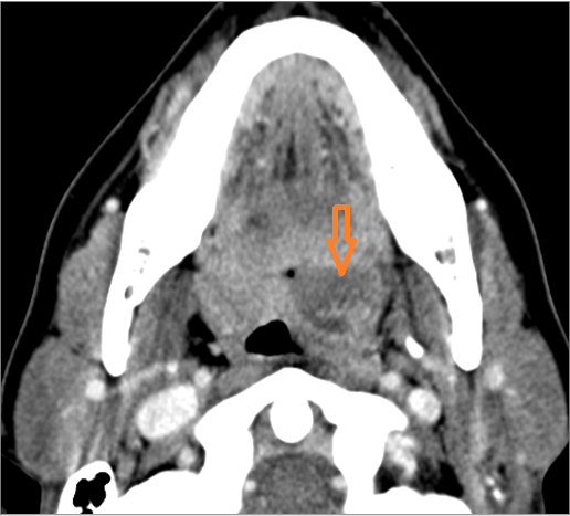
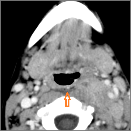
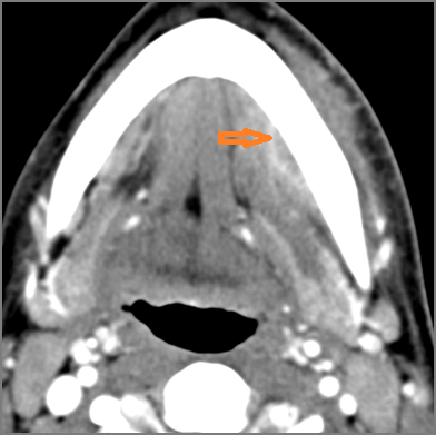
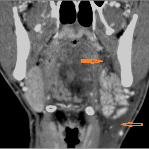
There is excessive enhancement or thickening of the fat or other soft tissues within or surrounding the buccal space, masticator space, floor of the mouth, submandibular space or the adjacent superficial fascia or subcutaneous fat and skin. |
Yes | NA |
|
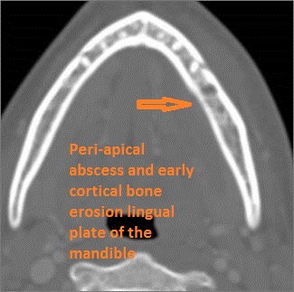
There is subperiosteal abscess or an abscess cavity adjacent to or involving the maxilla or mandible. |
No | NA |
|
There is endodontal or periodontal disease that might be causing cellulitis or abscess. |
Yes | NA |
|
There isedema/abscess within the fat of the adjacent parapharyngeal and retropharyngeal space. |
Yes | NA |
| Major Salivary Glands - Parotid Glands | Correct Answer | Your Answer |
|---|---|---|
|
The parotid glands are enlarged. |
No | NA |
|
The parotid glands do show abnormal enhancement. |
No | NA |
|
There are intraglandular cysts or sialocoeles. |
No | NA |
|
There is abscess within the parotid gland. |
No | NA |
|
The parotid ducts and the intraglandular ducts are dilated. |
No | NA |
|
The parotid ducts and the intraglandular ducts are with evidence of intraductal stones or other causes of obstruction. |
No | NA |
|
There is edema orabscess within the fat surrounding the parotid gland or of the adjacent masticator space or other spaces. |
No | NA |
|
There are abnormalities along the course of the facial nerve |
No | NA |
|
There are enlarged abnormal intraparotid, facial or posterior neck lymph nodes. |
No | NA |
| Major Salivary Glands - Submandibular Glands | Correct Answer | Your Answer |
|---|---|---|
|
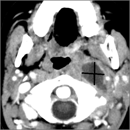
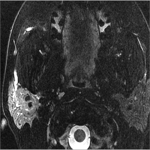
The submandibular glands are enlarged. |
Yes | NA |
|
The submandibular glands show abnormal enhancement. |
Yes | NA |
|
There areintraglandular cysts or sialocoeles. |
Yes | NA |
|
The submandibular ducts and the intraglandular ducts are dilated. |
No | NA |
|
The submandibular ducts and the intraglandular ducts are with evidence of intraductal stones other causes of obstruction. |
No | NA |
|
There is edema or abscess within the sublingual or submandibular space. |
Yes | NA |
|
The submandibular lymph nodes are abnormal by imaging criteria |
No | NA |
| Major Salivary Glands - Sublingual Glands | Correct Answer | Your Answer |
|---|---|---|
|
The sublingual glands are enlarged |
No | NA |
|
The sublingual glands do show abnormal enhancement. |
No | NA |
| Hypopharynx, Larynx, Deep Neck and Entire Retropharyngeal Space | Correct Answer | Your Answer |
|---|---|---|
|
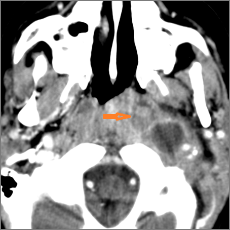
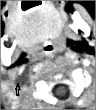
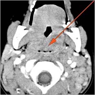
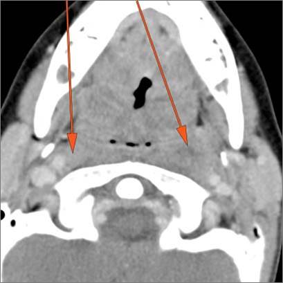
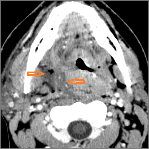
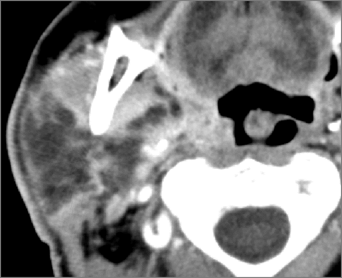
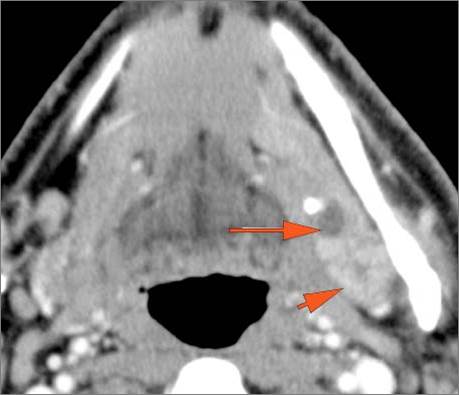
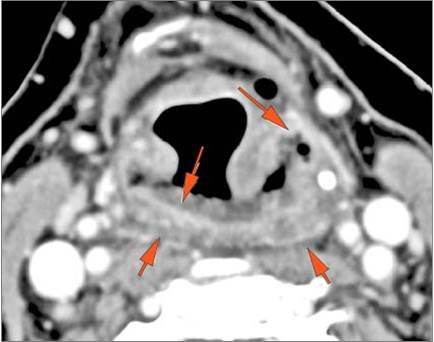
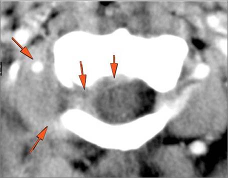
There isexcessive enhancement or thickening of the mucosa within in the hypopharynx, larynx or trachea. |
Yes | NA |
|
There is edema within in the hypopharynx, larynx or trachea. |
Yes | NA |
|
There isabscess within in the hypopharynx, larynx or trachea. |
No | NA |
|
There is edema within the adjacent deep neck, retropharyngeal and/or prevertebral spaces |
Yes | NA |
|
There isabscess within the adjacent deep neck, retropharyngeal and/or prevertebral spaces. |
No | NA |
| Prevertebral and Epidural Spaces | Correct Answer | Your Answer |
|---|---|---|
|
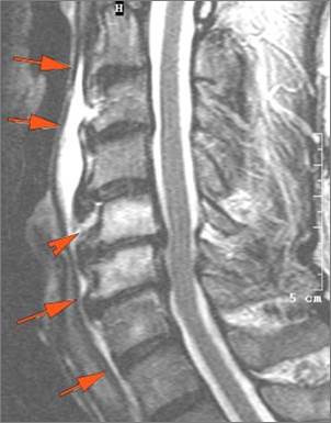
There isedema/abscess within in the prevertebral or paravertebral spaces. |
No | NA |
|
|
No | NA |
|
There iserosive process involving the disc spaces or other components of the spine. |
No | NA |
| Other Cervical Lymph Nodes | Correct Answer | Your Answer |
|---|---|---|
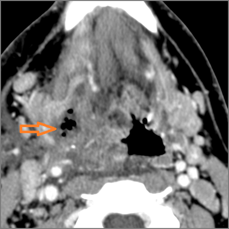
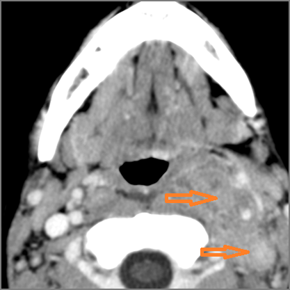
|
There is reactive cervical lymphadenopathy. |
Yes | NA |
|
There is suppurative cervical lymphadenopathy. |
No | NA |
|
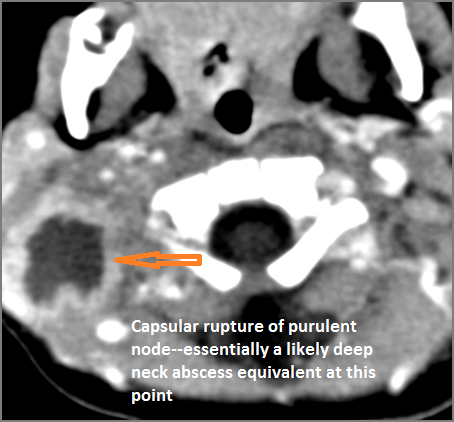
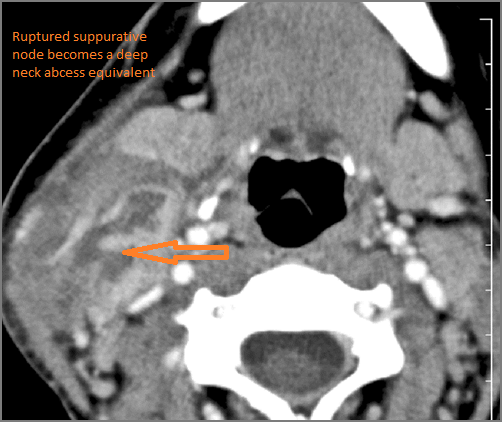
If there is suppurative cervical adenopathy thepurulent material outside the lymph node(s) capsule(s). |
No | NA |
| Vascular Findings | Correct Answer | Your Answer |
|---|---|---|
|
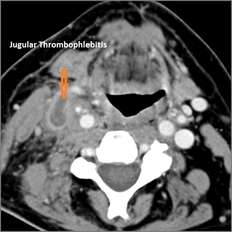
There is evidence of thrombus,thrombophlebitis or other occlusive or inflammatory process of the jugular vein or smaller venous tributaries. |
No | NA |
|
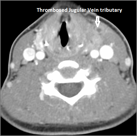
There is evidence of thrombus, thrombophlebitis or other occlusive or inflammatory processsmaller jugular venous tributaries. |
No | NA |
|
There is evidence of thrombus,inflammation of the common, external or internal carotid artery. |
No | NA |
|
There is evidence of acontained leakage from an arterial source. |
No | NA |
|
There is evidence ofactive extravasation from an arterial source |
No | NA |
| Upper Lung Zones and Mediastinum | Correct Answer | Your Answer |
|---|---|---|
|
The upper lung zones and mediastinum visualized are abnormal. |
No | NA |
| Other | Correct Answer | Your Answer |
|---|---|---|
|
Other significant abnormal findings are present |
No | NA |
Impression
Expert Answer
Pharyngitis and supraglottitis
